 Movie
Movie Controller

[English] 日本語
 Yorodumi
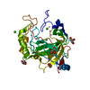
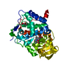
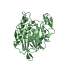
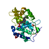
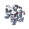
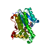
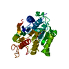
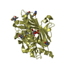
- PDB-1zhg: Crystal structure of Beta-Hydroxyacyl-Acyl Carrier Protein Dehydr... -
+ Open data

- Basic information
| Entry | Database: PDB / ID: 1zhg | ||||||
|---|---|---|---|---|---|---|---|
| Title | Crystal structure of Beta-Hydroxyacyl-Acyl Carrier Protein Dehydratase (FabZ) from Plasmodium falciparum | ||||||
 Components Components | beta hydroxyacyl-acyl carrier protein dehydratase | ||||||
 Keywords Keywords | LYASE / FabZ / Plasmodium falciparum / beta-hydroxyacyl acyl carrier protein dehydratase / hot dog fold | ||||||
| Function / homology |  Function and homology information Function and homology information3-hydroxyacyl-[acyl-carrier-protein] dehydratase / (3R)-hydroxyacyl-[acyl-carrier-protein] dehydratase activity / lipid A biosynthetic process / fatty acid biosynthetic process / membrane / cytoplasm Similarity search - Function | ||||||
| Biological species |  | ||||||
| Method |  X-RAY DIFFRACTION / MOLECULAR REPLACEMENT / Resolution: 2.4 Å X-RAY DIFFRACTION / MOLECULAR REPLACEMENT / Resolution: 2.4 Å | ||||||
 Authors Authors | Swarnamukhi, P.L. / Sharma, S.K. / Surolia, N. / Surolia, A. / Suguna, K. | ||||||
 Citation Citation | Journal: Febs Lett. / Year: 2006 Title: Crystal structure of dimeric FabZ of Plasmodium falciparum reveals conformational switching to active hexamers by peptide flips Authors: Swarnamukhi, P.L. / Sharma, S.K. / Bajaj, P. / Surolia, N. / Surolia, A. / Suguna, K. | ||||||
| History |
|
- Structure visualization
| Structure viewer | Molecule: MolmilJmol/JSmol |
|---|
- Downloads & links
-Download
| PDBx/mmCIF format | 1zhg.cif.gz | 64.6 KB | Display | PDBx/mmCIF format |
|---|---|---|---|---|
| PDB format | pdb1zhg.ent.gz | 47.5 KB | Display | PDB format |
| PDBx/mmJSON format | 1zhg.json.gz | Tree view | PDBx/mmJSON format | |
| Others |  Other downloads Other downloads |
-Validation report
| Arichive directory | https://data.pdbj.org/pub/pdb/validation_reports/zh/1zhgftp://data.pdbj.org/pub/pdb/validation_reports/zh/1zhg | HTTPS FTP |
|---|
-Related structure data
| Related structure data |  1u1zS S: Starting model for refinement |
|---|---|
| Similar structure data |


-Links
 PDBj
PDBj
- Assembly
| Deposited unit | 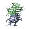
| ||||||||
|---|---|---|---|---|---|---|---|---|---|
| 1 |
| ||||||||
| Unit cell |
|
-Components
| #1: Protein | Mass: 15348.076 Da / Num. of mol.: 2 / Fragment: PfFabZ Source method: isolated from a genetically manipulated source Source: (gene. exp.) Gene: fabz / Plasmid: pET-28a(+) / Species (production host): Escherichia coli / Production host:  References: GenBank: 31322019, UniProt: Q965D7*PLUS, Lyases; Carbon-oxygen lyases; Hydro-lyases #2: Water | ChemComp-HOH / |  Mass: 18.015 Da / Num. of mol.: 95 / Source method: isolated from a natural source / Formula: H2O Mass: 18.015 Da / Num. of mol.: 95 / Source method: isolated from a natural source / Formula: H2O |
|---|
-Experimental details
-Experiment
| Experiment | Method: X-RAY DIFFRACTION / Number of used crystals: 1 |
|---|
- Sample preparation
| Crystal | Density Matthews: 2.1 Å3/Da / Density % sol: 41.8 % |
|---|---|
| Crystal grow | Temperature: 300 K / Method: micro batch (evoparation under oil) / pH: 4.5 Details: PEG 4000, sodium acetate, ammonium acetate, pH 4.5, Micro batch (Evoparation under oil), temperature 300K |
-Data collection
| Diffraction | Mean temperature: 100 K |
|---|---|
| Diffraction source | Source: ROTATING ANODE / Type: RIGAKU / Wavelength: 1.5418 Å |
| Detector | Type: MARRESEARCH / Detector: IMAGE PLATE / Date: Mar 9, 2005 |
| Radiation | Monochromator: osmic mirror / Protocol: SINGLE WAVELENGTH / Monochromatic (M) / Laue (L): M / Scattering type: x-ray |
| Radiation wavelength | Wavelength: 1.5418 Å / Relative weight: 1 |
| Reflection | Resolution: 2.4→20 Å / Num. all: 11482 / Num. obs: 11456 / % possible obs: 100 % / Observed criterion σ(F): 0 / Observed criterion σ(I): 0 / Redundancy: 6.3 % / Biso Wilson estimate: 36.9 Å2 / Rmerge(I) obs: 0.075 / Net I/σ(I): 20.7 |
| Reflection shell | Resolution: 2.4→2.53 Å / Redundancy: 6.2 % / Rmerge(I) obs: 0.418 / Mean I/σ(I) obs: 4.1 / Num. unique all: 1641 / % possible all: 100 |
- Processing
| Software |
| ||||||||||||||||||||||||||||||||||||
|---|---|---|---|---|---|---|---|---|---|---|---|---|---|---|---|---|---|---|---|---|---|---|---|---|---|---|---|---|---|---|---|---|---|---|---|---|---|
| Refinement | Method to determine structure: MOLECULAR REPLACEMENT Starting model: pdb entry 1u1z Resolution: 2.4→20 Å / Rfactor Rfree error: 0.01 / Data cutoff high absF: 1626990.62 / Data cutoff low absF: 0 / Isotropic thermal model: RESTRAINED / Cross valid method: THROUGHOUT / σ(F): 0 / σ(I): 0 / Stereochemistry target values: Engh & Huber
| ||||||||||||||||||||||||||||||||||||
| Solvent computation | Solvent model: FLAT MODEL / Bsol: 42.7966 Å2 / ksol: 0.320845 e/Å3 | ||||||||||||||||||||||||||||||||||||
| Displacement parameters | Biso mean: 42 Å2
| ||||||||||||||||||||||||||||||||||||
| Refine analyze |
| ||||||||||||||||||||||||||||||||||||
| Refinement step | Cycle: LAST / Resolution: 2.4→20 Å
| ||||||||||||||||||||||||||||||||||||
| Refine LS restraints |
| ||||||||||||||||||||||||||||||||||||
| LS refinement shell | Resolution: 2.4→2.55 Å / Rfactor Rfree error: 0.032 / Total num. of bins used: 6
| ||||||||||||||||||||||||||||||||||||
| Xplor file |
|
