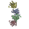
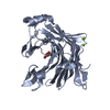
Deposited unit
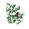
A: PHOSPHATIDYLINOSITOL TRANSFER PROTEIN ALPHA ISOFORM
B: PHOSPHATIDYLINOSITOL TRANSFER PROTEIN ALPHA ISOFORM
C: PHOSPHATIDYLINOSITOL TRANSFER PROTEIN ALPHA ISOFORM
D: PHOSPHATIDYLINOSITOL TRANSFER PROTEIN ALPHA ISOFORM
hetero molecules Summary Component details
Theoretical mass Number of molelcules Total (without water) 132,262 8 Polymers 128,918 4 Non-polymers 3,344 4 Water 0 0
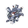
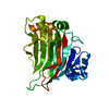
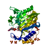
1
A: PHOSPHATIDYLINOSITOL TRANSFER PROTEIN ALPHA ISOFORM
hetero molecules Summary Component details Symmetry operations Calculated values
Theoretical mass Number of molelcules Total (without water) 33,066 2 Polymers 32,230 1 Non-polymers 836 1 Water 0
Type Name Symmetry operation Number identity operation 1_555 x,y,z 1
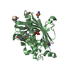
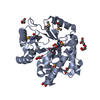
2
B: PHOSPHATIDYLINOSITOL TRANSFER PROTEIN ALPHA ISOFORM
hetero molecules Summary Component details Symmetry operations Calculated values
Theoretical mass Number of molelcules Total (without water) 33,066 2 Polymers 32,230 1 Non-polymers 836 1 Water 0
Type Name Symmetry operation Number identity operation 1_555 x,y,z 1
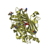
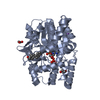
3
C: PHOSPHATIDYLINOSITOL TRANSFER PROTEIN ALPHA ISOFORM
hetero molecules Summary Component details Symmetry operations Calculated values
Theoretical mass Number of molelcules Total (without water) 33,066 2 Polymers 32,230 1 Non-polymers 836 1 Water 0
Type Name Symmetry operation Number identity operation 1_555 x,y,z 1
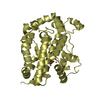
4
D: PHOSPHATIDYLINOSITOL TRANSFER PROTEIN ALPHA ISOFORM
hetero molecules Summary Component details Symmetry operations Calculated values
Theoretical mass Number of molelcules Total (without water) 33,066 2 Polymers 32,230 1 Non-polymers 836 1 Water 0
Type Name Symmetry operation Number identity operation 1_555 x,y,z 1
Unit cell Length a, b, c (Å) 83.262, 93.173, 161.732 Angle α, β, γ (deg.) 90.00, 90.00, 90.00 Int Tables number 19 Space group name H-M P21 21 21
Noncrystallographic symmetry (NCS) NCS domain ID Ens-ID Details (eV)1 1 A2 1 B3 1 C4 1 D1 2 A2 2 B3 2 C4 2 D
NCS domain segments Show large table (12 x 28) Hide large table Dom-ID Component-ID Ens-ID Beg auth comp-ID Beg label comp-ID End auth comp-ID End label comp-ID Refine code Auth asym-ID Label asym-ID Auth seq-ID Label seq-ID 1 1 1 MSEMSEASNASN2 AA1 - 45 1 - 45 2 1 1 MSEMSEASNASN2 BB1 - 45 1 - 45 3 1 1 MSEMSEASNASN2 CC1 - 45 1 - 45 4 1 1 MSEMSEASNASN2 DD1 - 45 1 - 45 1 2 1 GLUGLUTHRTHR6 AA46 - 58 46 - 58 2 2 1 GLUGLUTHRTHR6 BB46 - 58 46 - 58 3 2 1 GLUGLUTHRTHR6 CC46 - 58 46 - 58 4 2 1 GLUGLUTHRTHR6 DD46 - 58 46 - 58 1 3 1 TYRTYRILEILE2 AA57 - 98 57 - 98 2 3 1 TYRTYRILEILE2 BB57 - 98 57 - 98 3 3 1 TYRTYRILEILE2 CC57 - 98 57 - 98 4 3 1 TYRTYRILEILE2 DD57 - 98 57 - 98 1 4 1 THRTHRLYSLYS6 AA
 Movie
Movie Controller


 Open data
 Basic information
 Components
 Keywords
 Function and homology information
 HOMO SAPIENS (human)
 X-RAY DIFFRACTION /
  Authors
 Citation
 Structure visualization
 Downloads & links
 Other downloads


 PDBj
PDBj


 Assembly