 ムービー
ムービー コントローラー


+ データを開く

- 基本情報
基本情報
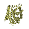
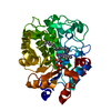
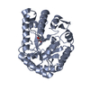
| 登録情報 | データベース: PDB / ID: 2aam | ||||||
|---|---|---|---|---|---|---|---|
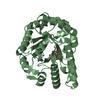
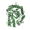
| タイトル | Crystal structure of a putative glycosidase (tm1410) from thermotoga maritima at 2.20 A resolution | ||||||
 要素 要素 | Hypothetical protein TM1410 | ||||||
 キーワード キーワード | HYDROLASE / Structural genomics / Joint Center for Structural Genomics / JCSG / Protein Structure Initiative / PSI-2 | ||||||
| 機能・相同性 |  機能・相同性情報 機能・相同性情報Extracellular protein / Hypothetical protein TM1410-related / Glycoside-hydrolase family GH114, TIM-barrel domain / Glycoside-hydrolase family GH114 / Aldolase class I / Aldolase-type TIM barrel / Glycoside hydrolase superfamily / TIM Barrel / Alpha-Beta Barrel / Alpha Beta 類似検索 - ドメイン・相同性 | ||||||
| 生物種 |   Thermotoga maritima (バクテリア) Thermotoga maritima (バクテリア) | ||||||
| 手法 |  X線回折 / シンクロトロン / 多波長異常分散 / 解像度: 2.2 Å X線回折 / シンクロトロン / 多波長異常分散 / 解像度: 2.2 Å | ||||||
 データ登録者 データ登録者 | Joint Center for Structural Genomics (JCSG) | ||||||
 引用 引用 | ジャーナル: To be published タイトル: Crystal structure of hypothetical protein (tm1410) from THERMOTOGA MARITIMA at 2.20 A resolution 著者: Joint Center for Structural Genomics (JCSG) | ||||||
| 履歴 |
|
- 構造の表示
構造の表示
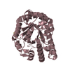
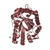
| 構造ビューア | 分子: MolmilJmol/JSmol |
|---|
- ダウンロードとリンク
-ダウンロード
| PDBx/mmCIF形式 | 2aam.cif.gz | 382 KB | 表示 | PDBx/mmCIF形式 |
|---|---|---|---|---|
| PDB形式 | pdb2aam.ent.gz | 311.6 KB | 表示 | PDB形式 |
| PDBx/mmJSON形式 | 2aam.json.gz | ツリー表示 | PDBx/mmJSON形式 | |
| その他 |  その他のダウンロード その他のダウンロード |
-検証レポート
| アーカイブディレクトリ | https://data.pdbj.org/pub/pdb/validation_reports/aa/2aamftp://data.pdbj.org/pub/pdb/validation_reports/aa/2aam | HTTPS FTP |
|---|
-関連構造データ
| 類似構造データ | |
|---|---|
| その他のデータベース |


-リンク
 PDBj
PDBj

- 集合体
集合体
| 登録構造単位 | 
| ||||||||||||||||||||||||||||||||||||||||||
|---|---|---|---|---|---|---|---|---|---|---|---|---|---|---|---|---|---|---|---|---|---|---|---|---|---|---|---|---|---|---|---|---|---|---|---|---|---|---|---|---|---|---|---|
| 1 | 
| ||||||||||||||||||||||||||||||||||||||||||
| 2 | 
| ||||||||||||||||||||||||||||||||||||||||||
| 3 | 
| ||||||||||||||||||||||||||||||||||||||||||
| 4 | 
| ||||||||||||||||||||||||||||||||||||||||||
| 5 | 
| ||||||||||||||||||||||||||||||||||||||||||
| 6 | 
| ||||||||||||||||||||||||||||||||||||||||||
| 単位格子 |
| ||||||||||||||||||||||||||||||||||||||||||
| 非結晶学的対称性 (NCS) | NCSドメイン:
NCSドメイン領域: Component-ID: 1 / Ens-ID: 1 / Beg auth comp-ID: TRP / Beg label comp-ID: TRP / End auth comp-ID: PRO / End label comp-ID: PRO / Refine code: 4 / Auth seq-ID: 30 - 310 / Label seq-ID: 16 - 296
|
-要素
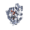
| #1: タンパク質 | 分子量: 36579.688 Da / 分子数: 6 / 由来タイプ: 組換発現 由来: (組換発現) Thermotoga maritima (バクテリア)遺伝子: tm1410 / プラスミド: DL41 / 発現宿主: #2: 化合物 | ChemComp-UNL / 分子数: 6 / 由来タイプ: 合成 #3: 化合物 | ChemComp-GOL /   分子量: 92.094 Da / 分子数: 37 / 由来タイプ: 合成 / 式: C3H8O3 分子量: 92.094 Da / 分子数: 37 / 由来タイプ: 合成 / 式: C3H8O3#4: 水 | ChemComp-HOH / |  分子量: 18.015 Da / 分子数: 926 / 由来タイプ: 天然 / 式: H2O 分子量: 18.015 Da / 分子数: 926 / 由来タイプ: 天然 / 式: H2OHas protein modification | Y | |
|---|
-実験情報
-実験
| 実験 | 手法: X線回折 / 使用した結晶の数: 1 |
|---|
- 試料調製
試料調製
| 結晶 | マシュー密度: 6.43 Å3/Da / 溶媒含有率: 80.71 % |
|---|---|
| 結晶化 | 温度: 277 K 手法: 蒸気拡散法, シッティングドロップ法, nanodrop pH: 9.5 詳細: 25.0% PEG-300, 10.0% Glycerol, 5.0% PEG-8000, 0.1M CHES pH 9.5 , VAPOR DIFFUSION,SITTING DROP,NANODROP, temperature 277K |
-データ収集
| 回折 | 平均測定温度: 100 K | |||||||||||||||||||||||||||||||||||||||||||||||||||||||||||||||||||||||||||||
|---|---|---|---|---|---|---|---|---|---|---|---|---|---|---|---|---|---|---|---|---|---|---|---|---|---|---|---|---|---|---|---|---|---|---|---|---|---|---|---|---|---|---|---|---|---|---|---|---|---|---|---|---|---|---|---|---|---|---|---|---|---|---|---|---|---|---|---|---|---|---|---|---|---|---|---|---|---|---|
| 放射光源 | 由来: シンクロトロン / サイト: SSRL  / ビームライン: BL11-1 / 波長: 0.979170, 0.918370 / ビームライン: BL11-1 / 波長: 0.979170, 0.918370 | |||||||||||||||||||||||||||||||||||||||||||||||||||||||||||||||||||||||||||||
| 検出器 | タイプ: ADSC QUANTUM 315 / 検出器: CCD / 日付: 2005年5月1日 / 詳細: flat mirror | |||||||||||||||||||||||||||||||||||||||||||||||||||||||||||||||||||||||||||||
| 放射 | プロトコル: MAD / 単色(M)・ラウエ(L): M / 散乱光タイプ: x-ray | |||||||||||||||||||||||||||||||||||||||||||||||||||||||||||||||||||||||||||||
| 放射波長 |
| |||||||||||||||||||||||||||||||||||||||||||||||||||||||||||||||||||||||||||||
| 反射 | 解像度: 2.2→28.59 Å / Num. obs: 263407 / % possible obs: 90.2 % / Rmerge(I) obs: 0.093 / Net I/σ(I): 8.15 | |||||||||||||||||||||||||||||||||||||||||||||||||||||||||||||||||||||||||||||
| 反射 シェル |
|
-位相決定
| 位相決定 | 手法: 多波長異常分散 |
|---|
- 解析
解析
| ソフトウェア |
| |||||||||||||||||||||||||||||||||||||||||||||||||||||||||||||||||||||||||||||||||||||||||||||||||||||||||||||||||||||||||||||||||||||||||||||||||||||||||||||||||||||||||||||||
|---|---|---|---|---|---|---|---|---|---|---|---|---|---|---|---|---|---|---|---|---|---|---|---|---|---|---|---|---|---|---|---|---|---|---|---|---|---|---|---|---|---|---|---|---|---|---|---|---|---|---|---|---|---|---|---|---|---|---|---|---|---|---|---|---|---|---|---|---|---|---|---|---|---|---|---|---|---|---|---|---|---|---|---|---|---|---|---|---|---|---|---|---|---|---|---|---|---|---|---|---|---|---|---|---|---|---|---|---|---|---|---|---|---|---|---|---|---|---|---|---|---|---|---|---|---|---|---|---|---|---|---|---|---|---|---|---|---|---|---|---|---|---|---|---|---|---|---|---|---|---|---|---|---|---|---|---|---|---|---|---|---|---|---|---|---|---|---|---|---|---|---|---|---|---|---|---|
| 精密化 | 構造決定の手法: 多波長異常分散 / 解像度: 2.2→28.59 Å / Cor.coef. Fo:Fc: 0.919 / Cor.coef. Fo:Fc free: 0.902 / SU B: 7.963 / SU ML: 0.105 / TLS residual ADP flag: LIKELY RESIDUAL / 交差検証法: THROUGHOUT / ESU R: 0.134 / ESU R Free: 0.13 立体化学のターゲット値: MAXIMUM LIKELIHOOD WITH PHASES 詳細: 1. HYDROGENS HAVE BEEN ADDED IN THE RIDING POSITIONS 2. ELECTRON DENSITY IS DISORDERED AT THE N AND C-TERMINI, THEREFORE, THE STRUCTURE WAS NOT MODELED IN THESE REGIONS 3. ELECTRON DENSITY ...詳細: 1. HYDROGENS HAVE BEEN ADDED IN THE RIDING POSITIONS 2. ELECTRON DENSITY IS DISORDERED AT THE N AND C-TERMINI, THEREFORE, THE STRUCTURE WAS NOT MODELED IN THESE REGIONS 3. ELECTRON DENSITY NEAR THE SIDECHAIN OF TYR 98 ON EACH SUBUNIT INDICATES A BOUND LIGAND. BASED ON STRUCTURAL HOMOLOGS, IT IS BELIEVED THAT THE UNKNOWN LIGAND CONTAINS A MODIFIED PYRANOSE RING.
| |||||||||||||||||||||||||||||||||||||||||||||||||||||||||||||||||||||||||||||||||||||||||||||||||||||||||||||||||||||||||||||||||||||||||||||||||||||||||||||||||||||||||||||||
| 溶媒の処理 | イオンプローブ半径: 0.8 Å / 減衰半径: 0.8 Å / VDWプローブ半径: 1.2 Å / 溶媒モデル: BABINET MODEL WITH MASK | |||||||||||||||||||||||||||||||||||||||||||||||||||||||||||||||||||||||||||||||||||||||||||||||||||||||||||||||||||||||||||||||||||||||||||||||||||||||||||||||||||||||||||||||
| 原子変位パラメータ | Biso mean: 37.156 Å2
| |||||||||||||||||||||||||||||||||||||||||||||||||||||||||||||||||||||||||||||||||||||||||||||||||||||||||||||||||||||||||||||||||||||||||||||||||||||||||||||||||||||||||||||||
| 精密化ステップ | サイクル: LAST / 解像度: 2.2→28.59 Å
| |||||||||||||||||||||||||||||||||||||||||||||||||||||||||||||||||||||||||||||||||||||||||||||||||||||||||||||||||||||||||||||||||||||||||||||||||||||||||||||||||||||||||||||||
| 拘束条件 |
| |||||||||||||||||||||||||||||||||||||||||||||||||||||||||||||||||||||||||||||||||||||||||||||||||||||||||||||||||||||||||||||||||||||||||||||||||||||||||||||||||||||||||||||||
| Refine LS restraints NCS | Ens-ID: 1 / 数: 4262 / Refine-ID: X-RAY DIFFRACTION
| |||||||||||||||||||||||||||||||||||||||||||||||||||||||||||||||||||||||||||||||||||||||||||||||||||||||||||||||||||||||||||||||||||||||||||||||||||||||||||||||||||||||||||||||
| LS精密化 シェル | 解像度: 2.2→2.258 Å / Total num. of bins used: 20
| |||||||||||||||||||||||||||||||||||||||||||||||||||||||||||||||||||||||||||||||||||||||||||||||||||||||||||||||||||||||||||||||||||||||||||||||||||||||||||||||||||||||||||||||
| 精密化 TLS | 手法: refined / Refine-ID: X-RAY DIFFRACTION
| |||||||||||||||||||||||||||||||||||||||||||||||||||||||||||||||||||||||||||||||||||||||||||||||||||||||||||||||||||||||||||||||||||||||||||||||||||||||||||||||||||||||||||||||
| 精密化 TLSグループ | Refine-ID: X-RAY DIFFRACTION / Selection: all
|
