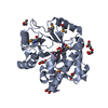
[English] 日本語
Yorodumi
- PDB-6dno: Crystal structure of Protein Phosphatase 1 (PP1) bound to the mus... -

+
Open data


ID or keywords:

Loading...

-
Basic information

Entry
Database: PDB / ID: 6dno
TitleCrystal structure of Protein Phosphatase 1 (PP1) bound to the muscle glycogen-targeting subunit (Gm)
Components
  • Microcystin-LR
  • Protein phosphatase 1 regulatory subunit 3A
  • Serine/threonine-protein phosphatase PP1-alpha catalytic subunit
KeywordsSIGNALING PROTEIN / Inhibitor / complex / HYDROLASE / HYDROLASE-HYDROLASE INHIBITOR complex
Function / homology
Function and homology information


protein phosphatase type 1 complex / regulation of glycogen catabolic process / PTW/PP1 phosphatase complex / glycogen granule / regulation of glycogen biosynthetic process / protein phosphatase 1 binding / cadherin binding involved in cell-cell adhesion / positive regulation of extrinsic apoptotic signaling pathway in absence of ligand / regulation of canonical Wnt signaling pathway / regulation of translational initiation ...protein phosphatase type 1 complex / regulation of glycogen catabolic process / PTW/PP1 phosphatase complex / glycogen granule / regulation of glycogen biosynthetic process / protein phosphatase 1 binding / cadherin binding involved in cell-cell adhesion / positive regulation of extrinsic apoptotic signaling pathway in absence of ligand / regulation of canonical Wnt signaling pathway / regulation of translational initiation / myosin phosphatase activity / protein serine/threonine phosphatase activity / branching morphogenesis of an epithelial tube / glycogen metabolic process / protein-serine/threonine phosphatase / entrainment of circadian clock by photoperiod / Triglyceride catabolism / phosphatase activity / phosphoprotein phosphatase activity / DARPP-32 events / ribonucleoprotein complex binding / dephosphorylation / Downregulation of TGF-beta receptor signaling / protein dephosphorylation / adherens junction / response to lead ion / lung development / circadian regulation of gene expression / regulation of circadian rhythm / Circadian Clock / presynapse / perikaryon / dendritic spine / cell cycle / cell division / glutamatergic synapse / nucleolus / extracellular exosome / nucleoplasm / membrane / metal ion binding / nucleus / plasma membrane / cytosol / cytoplasm
Similarity search - Function
CBM21 (carbohydrate binding type-21) domain / CBM21 domain superfamily / Carbohydrate/starch-binding module (family 21) / CBM21 (carbohydrate binding type-21) domain profile. / Serine-threonine protein phosphatase, N-terminal / Serine-threonine protein phosphatase N-terminal domain / Serine/threonine specific protein phosphatases signature. / Protein phosphatase 2A homologues, catalytic domain. / Serine/threonine-specific protein phosphatase/bis(5-nucleosyl)-tetraphosphatase / Metallo-dependent phosphatases ...CBM21 (carbohydrate binding type-21) domain / CBM21 domain superfamily / Carbohydrate/starch-binding module (family 21) / CBM21 (carbohydrate binding type-21) domain profile. / Serine-threonine protein phosphatase, N-terminal / Serine-threonine protein phosphatase N-terminal domain / Serine/threonine specific protein phosphatases signature. / Protein phosphatase 2A homologues, catalytic domain. / Serine/threonine-specific protein phosphatase/bis(5-nucleosyl)-tetraphosphatase / Metallo-dependent phosphatases / Purple Acid Phosphatase; chain A, domain 2 / Calcineurin-like phosphoesterase domain, ApaH type / Calcineurin-like phosphoesterase / Metallo-dependent phosphatase-like / 4-Layer Sandwich / Alpha Beta
Similarity search - Domain/homology
Microcystin LR / Serine/threonine-protein phosphatase PP1-alpha catalytic subunit / Protein phosphatase 1 regulatory subunit 3A
Similarity search - Component
Biological speciesHomo sapiens (human)
Oryctolagus cuniculus (rabbit)
synthetic construct (others)
MethodX-RAY DIFFRACTION / SYNCHROTRON / MOLECULAR REPLACEMENT / Resolution: 1.45 Å
AuthorsChoy, M.S. / Kumar, G.S. / Peti, W. / Page, R.
Funding support United States, 2items
OrganizationGrant numberCountry
National Institutes of Health/National Institute on Drug Abuse (NIH/NIDA)GM098482 United States
National Institutes of Health/National Institute on Drug Abuse (NIH/NIDA)NS091336 United States
CitationJournal: Sci Adv / Year: 2018
Title: Identification of the substrate recruitment mechanism of the muscle glycogen protein phosphatase 1 holoenzyme.
Authors: Kumar, G.S. / Choy, M.S. / Koveal, D.M. / Lorinsky, M.K. / Lyons, S.P. / Kettenbach, A.N. / Page, R. / Peti, W.
History
DepositionJun 7, 2018Deposition site: RCSB / Processing site: RCSB
Revision 1.0Jan 23, 2019Provider: repository / Type: Initial release
Revision 1.1Dec 11, 2019Group: Author supporting evidence / Category: pdbx_audit_support / Item: _pdbx_audit_support.funding_organization
Revision 1.2Oct 11, 2023Group: Data collection / Database references ...Data collection / Database references / Derived calculations / Refinement description
Category: chem_comp_atom / chem_comp_bond ...chem_comp_atom / chem_comp_bond / database_2 / pdbx_initial_refinement_model / struct_conn
Item: _database_2.pdbx_DOI / _database_2.pdbx_database_accession ..._database_2.pdbx_DOI / _database_2.pdbx_database_accession / _struct_conn.pdbx_dist_value / _struct_conn.ptnr1_label_atom_id / _struct_conn.ptnr2_auth_comp_id / _struct_conn.ptnr2_auth_seq_id / _struct_conn.ptnr2_label_atom_id / _struct_conn.ptnr2_label_comp_id / _struct_conn.ptnr2_label_seq_id
Revision 2.0Nov 15, 2023Group: Atomic model / Data collection / Derived calculations
Category: atom_site / chem_comp_atom ...atom_site / chem_comp_atom / chem_comp_bond / pdbx_validate_main_chain_plane / pdbx_validate_peptide_omega / pdbx_validate_rmsd_bond / struct_conn
Item: _atom_site.auth_atom_id / _atom_site.label_atom_id ..._atom_site.auth_atom_id / _atom_site.label_atom_id / _chem_comp_atom.atom_id / _chem_comp_bond.atom_id_1 / _chem_comp_bond.atom_id_2 / _struct_conn.ptnr1_label_atom_id / _struct_conn.ptnr2_label_atom_id

-
Structure visualization

Structure viewerMolecule:
MolmilJmol/JSmol

Downloads & links

-
Assembly

Deposited unit
A: Serine/threonine-protein phosphatase PP1-alpha catalytic subunit
B: Protein phosphatase 1 regulatory subunit 3A
C: Microcystin-LR


Theoretical massNumber of molelcules
Total (without water)38,6293
Polymers38,6293
Non-polymers00
Water2,648147
1


  • Idetical with deposited unit
  • defined by author&software
  • Evidence: gel filtration
TypeNameSymmetry operationNumber
identity operation1_555x,y,z1
Buried area3460 Å2
ΔGint-17 kcal/mol
Surface area13220 Å2
MethodPISA
Unit cell
Length a, b, c (Å)60.476, 65.744, 160.355
Angle α, β, γ (deg.)90.00, 90.00, 90.00
Int Tables number20
Space group name H-MC2221
Components on special symmetry positions
IDModelComponents
11B-102-

HOH

-
Components

#1: Protein Serine/threonine-protein phosphatase PP1-alpha catalytic subunit / PP-1A


Mass: 34162.148 Da / Num. of mol.: 1 / Fragment: residues 7-300
Source method: isolated from a genetically manipulated source
Source: (gene. exp.) Homo sapiens (human) / Gene: PPP1CA, PPP1A / Production host: Escherichia coli (E. coli)
References: UniProt: P62136, protein-serine/threonine phosphatase
#2: Protein/peptide Protein phosphatase 1 regulatory subunit 3A / Protein phosphatase 1 glycogen-associated regulatory subunit / Protein phosphatase type-1 glycogen ...Protein phosphatase 1 glycogen-associated regulatory subunit / Protein phosphatase type-1 glycogen targeting subunit / RG1


Mass: 3452.802 Da / Num. of mol.: 1 / Fragment: residues 64-93 / Source method: obtained synthetically / Source: (synth.) Oryctolagus cuniculus (rabbit) / References: UniProt: Q00756
#3: Protein/peptide Microcystin-LR / / Microcystin-LR


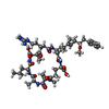
Type: Oligopeptide / Class: Toxin / Mass: 1014.195 Da / Num. of mol.: 1 / Source method: obtained synthetically / Source: (synth.) synthetic construct (others) / References: Microcystin LR
#4: Water ChemComp-HOH / water / Water


Mass: 18.015 Da / Num. of mol.: 147 / Source method: isolated from a natural source / Formula: H2O

-
Experimental details

-
Experiment

ExperimentMethod: X-RAY DIFFRACTION / Number of used crystals: 1

-
Sample preparation

CrystalDensity Matthews: 2.12 Å3/Da / Density % sol: 41.95 %
Crystal growTemperature: 277 K / Method: vapor diffusion / pH: 4.6
Details: 0.4 M magnesium formate dihydrate, 0.1 M Sodium Acetate trihydrate

-
Data collection

DiffractionMean temperature: 100 K
Diffraction sourceSource: SYNCHROTRON / Site: SSRL / Beamline: BL12-2 / Wavelength: 0.97945 Å
DetectorType: DECTRIS PILATUS3 6M / Detector: PIXEL / Date: Feb 4, 2016
RadiationProtocol: SINGLE WAVELENGTH / Monochromatic (M) / Laue (L): M / Scattering type: x-ray
Radiation wavelengthWavelength: 0.97945 Å / Relative weight: 1
ReflectionResolution: 1.45→38.9 Å / Num. obs: 56091 / % possible obs: 98.5 % / Redundancy: 5.1 % / CC1/2: 0.998 / Rmerge(I) obs: 0.061 / Net I/σ(I): 12.2
Reflection shellResolution: 1.45→1.47 Å / Redundancy: 2.9 % / Rmerge(I) obs: 1.147 / Mean I/σ(I) obs: 0.9 / CC1/2: 0.337 / % possible all: 72.9

-
Processing

Software
NameVersionClassification
PHENIX(1.13_2998)refinement
XDSoct 15, 2015data reduction
Aimless0.5.17data scaling
PHENIX2.5.6phasing
RefinementMethod to determine structure: MOLECULAR REPLACEMENT
Starting model: 4mov
Resolution: 1.45→38.9 Å / SU ML: 0.18 / Cross valid method: FREE R-VALUE / σ(F): 1.34 / Phase error: 22.39
RfactorNum. reflection% reflectionSelection details
Rfree0.2096 2680 4.78 %random selection
Rwork0.1859 ---
obs0.187 56035 98.39 %-
Solvent computationShrinkage radii: 0.9 Å / VDW probe radii: 1.11 Å
Refinement stepCycle: LAST / Resolution: 1.45→38.9 Å
ProteinNucleic acidLigandSolventTotal
Num. atoms2547 0 71 147 2765
Refine LS restraints
Refine-IDTypeDev idealNumber
X-RAY DIFFRACTIONf_bond_d0.0052698
X-RAY DIFFRACTIONf_angle_d0.9633650
X-RAY DIFFRACTIONf_dihedral_angle_d21.863988
X-RAY DIFFRACTIONf_chiral_restr0.081390
X-RAY DIFFRACTIONf_plane_restr0.004478
LS refinement shell
Resolution (Å)Rfactor RfreeNum. reflection RfreeRfactor RworkNum. reflection RworkRefine-ID% reflection obs (%)
1.4504-1.47670.36091150.33032094X-RAY DIFFRACTION74
1.4767-1.50510.33311450.29362785X-RAY DIFFRACTION99
1.5051-1.53590.30191430.26792835X-RAY DIFFRACTION100
1.5359-1.56930.27441390.2642793X-RAY DIFFRACTION100
1.5693-1.60580.25411350.24952840X-RAY DIFFRACTION100
1.6058-1.64590.24971480.24082839X-RAY DIFFRACTION100
1.6459-1.69040.23511340.22732810X-RAY DIFFRACTION100
1.6904-1.74020.25251250.21552834X-RAY DIFFRACTION100
1.7402-1.79630.23681500.21172821X-RAY DIFFRACTION100
1.7963-1.86050.23191590.20722842X-RAY DIFFRACTION100
1.8605-1.9350.25291430.20232837X-RAY DIFFRACTION100
1.935-2.02310.18951440.19992826X-RAY DIFFRACTION100
2.0231-2.12970.2121510.18072828X-RAY DIFFRACTION100
2.1297-2.26320.21961640.18572818X-RAY DIFFRACTION100
2.2632-2.43790.2041300.17852879X-RAY DIFFRACTION100
2.4379-2.68320.2131280.18812864X-RAY DIFFRACTION99
2.6832-3.07130.19241140.18092893X-RAY DIFFRACTION99
3.0713-3.86890.18821610.16262905X-RAY DIFFRACTION100
3.8689-38.9290.17791520.15433012X-RAY DIFFRACTION99

+
About Yorodumi

-
News

-
Feb 9, 2022. New format data for meta-information of EMDB entries

New format data for meta-information of EMDB entries

  • Version 3 of the EMDB header file is now the official format.
  • The previous official version 1.9 will be removed from the archive.

Related info.:EMDB header

External links:wwPDB to switch to version 3 of the EMDB data model

-
Aug 12, 2020. Covid-19 info

Covid-19 info

URL: https://pdbj.org/emnavi/covid19.php

New page: Covid-19 featured information page in EM Navigator.

Related info.:Covid-19 info / Mar 5, 2020. Novel coronavirus structure data

+
Mar 5, 2020. Novel coronavirus structure data

Novel coronavirus structure data

Related info.:Yorodumi Speices / Aug 12, 2020. Covid-19 info

External links:COVID-19 featured content - PDBj / Molecule of the Month (242):Coronavirus Proteases

+
Jan 31, 2019. EMDB accession codes are about to change! (news from PDBe EMDB page)

EMDB accession codes are about to change! (news from PDBe EMDB page)

  • The allocation of 4 digits for EMDB accession codes will soon come to an end. Whilst these codes will remain in use, new EMDB accession codes will include an additional digit and will expand incrementally as the available range of codes is exhausted. The current 4-digit format prefixed with “EMD-” (i.e. EMD-XXXX) will advance to a 5-digit format (i.e. EMD-XXXXX), and so on. It is currently estimated that the 4-digit codes will be depleted around Spring 2019, at which point the 5-digit format will come into force.
  • The EM Navigator/Yorodumi systems omit the EMD- prefix.

Related info.:Q: What is EMD? / ID/Accession-code notation in Yorodumi/EM Navigator

External links:EMDB Accession Codes are Changing Soon! / Contact to PDBj

+
Jul 12, 2017. Major update of PDB

Major update of PDB

  • wwPDB released updated PDB data conforming to the new PDBx/mmCIF dictionary.
  • This is a major update changing the version number from 4 to 5, and with Remediation, in which all the entries are updated.
  • In this update, many items about electron microscopy experimental information are reorganized (e.g. em_software).
  • Now, EM Navigator and Yorodumi are based on the updated data.

External links:wwPDB Remediation / Enriched Model Files Conforming to OneDep Data Standards Now Available in the PDB FTP Archive

-
Yorodumi

Thousand views of thousand structures

  • Yorodumi is a browser for structure data from EMDB, PDB, SASBDB, etc.
  • This page is also the successor to EM Navigator detail page, and also detail information page/front-end page for Omokage search.
  • The word "yorodu" (or yorozu) is an old Japanese word meaning "ten thousand". "mi" (miru) is to see.

Related info.:EMDB / PDB / SASBDB / Comparison of 3 databanks / Yorodumi Search / Aug 31, 2016. New EM Navigator & Yorodumi / Yorodumi Papers / Jmol/JSmol / Function and homology information / Changes in new EM Navigator and Yorodumi

Read more