 Movie
Movie Controller

[English] 日本語
 Yorodumi
- PDB-1qfj: CRYSTAL STRUCTURE OF NAD(P)H:FLAVIN OXIDOREDUCTASE FROM ESCHERICH... -
+ Open data

- Basic information
| Entry | Database: PDB / ID: 1qfj | ||||||
|---|---|---|---|---|---|---|---|
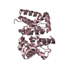
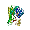
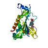
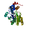
| Title | CRYSTAL STRUCTURE OF NAD(P)H:FLAVIN OXIDOREDUCTASE FROM ESCHERICHIA COLI | ||||||
 Components Components | PROTEIN (FLAVIN REDUCTASE) | ||||||
 Keywords Keywords | OXIDOREDUCTASE / RIBOFLAVIN / FLAVIN REDUCTASE / FERREDOXIN REDUCTASE SUPERFAMILY | ||||||
| Function / homology |  Function and homology information Function and homology informationriboflavin reductase [NAD(P)H] / riboflavin reductase [NAD(P)H] activity / : / riboflavin reductase (NADPH) activity / protein repair / monoatomic ion transport / iron ion transport / enzyme activator activity / response to oxidative stress / oxidoreductase activity / cytosol Similarity search - Function | ||||||
| Biological species |  | ||||||
| Method |  X-RAY DIFFRACTION / SYNCHROTRON / MAD / Resolution: 2.2 Å X-RAY DIFFRACTION / SYNCHROTRON / MAD / Resolution: 2.2 Å | ||||||
 Authors Authors | Ingelman, M. / Ramaswamy, S. / Niviere, V. / Fontecave, M. / Eklund, H. | ||||||
 Citation Citation | Journal: Biochemistry / Year: 1999 Title: Crystal structure of NAD(P)H:flavin oxidoreductase from Escherichia coli. Authors: Ingelman, M. / Ramaswamy, S. / Niviere, V. / Fontecave, M. / Eklund, H. | ||||||
| History |
|
- Structure visualization
| Structure viewer | Molecule: MolmilJmol/JSmol |
|---|
- Downloads & links
-Download
| PDBx/mmCIF format | 1qfj.cif.gz | 189.7 KB | Display | PDBx/mmCIF format |
|---|---|---|---|---|
| PDB format | pdb1qfj.ent.gz | 154.5 KB | Display | PDB format |
| PDBx/mmJSON format | 1qfj.json.gz | Tree view | PDBx/mmJSON format | |
| Others |  Other downloads Other downloads |
-Validation report
| Arichive directory | https://data.pdbj.org/pub/pdb/validation_reports/qf/1qfjftp://data.pdbj.org/pub/pdb/validation_reports/qf/1qfj | HTTPS FTP |
|---|
-Related structure data
| Similar structure data |
|---|


-Links
 PDBj
PDBj

- Assembly
| Deposited unit | 
| ||||||||||||||||
|---|---|---|---|---|---|---|---|---|---|---|---|---|---|---|---|---|---|
| 1 | 
| ||||||||||||||||
| 2 | 
| ||||||||||||||||
| 3 | 
| ||||||||||||||||
| 4 | 
| ||||||||||||||||
| Unit cell |
| ||||||||||||||||
| Noncrystallographic symmetry (NCS) | NCS oper:
|
-Components
| #1: Protein | Mass: 26143.791 Da / Num. of mol.: 4 Source method: isolated from a genetically manipulated source Details: DISULFIDE LINKS BETWEEN MOLECULES A AND D AS WELL AS B AND C RESPECTIVELY Source: (gene. exp.) #2: Chemical | ChemComp-GOL /   Mass: 92.094 Da / Num. of mol.: 4 / Source method: obtained synthetically / Formula: C3H8O3 Mass: 92.094 Da / Num. of mol.: 4 / Source method: obtained synthetically / Formula: C3H8O3#3: Water | ChemComp-HOH / |  Mass: 18.015 Da / Num. of mol.: 304 / Source method: isolated from a natural source / Formula: H2O Mass: 18.015 Da / Num. of mol.: 304 / Source method: isolated from a natural source / Formula: H2OHas protein modification | Y | |
|---|
-Experimental details
-Experiment
| Experiment | Method: X-RAY DIFFRACTION / Number of used crystals: 1 |
|---|
- Sample preparation
| Crystal | Density Matthews: 2.7 Å3/Da / Density % sol: 51 % | ||||||||||||||||||||||||||||||
|---|---|---|---|---|---|---|---|---|---|---|---|---|---|---|---|---|---|---|---|---|---|---|---|---|---|---|---|---|---|---|---|
| Crystal grow | pH: 6.5 Details: 15 % PEG 4000, 0.5 M NACL, 100 MM BIS-TRIS PROPANE PH 6.5 | ||||||||||||||||||||||||||||||
| Crystal grow | *PLUS Temperature: 4 ℃ / Method: vapor diffusion | ||||||||||||||||||||||||||||||
| Components of the solutions | *PLUS
|
-Data collection
| Diffraction | Mean temperature: 100 K |
|---|---|
| Diffraction source | Source: SYNCHROTRON / Site: EMBL/DESY, HAMBURG  / Beamline: BW7B / Wavelength: 0.864 / Beamline: BW7B / Wavelength: 0.864 |
| Detector | Type: MARRESEARCH / Detector: IMAGE PLATE / Date: Jun 1, 1995 |
| Radiation | Protocol: SINGLE WAVELENGTH / Monochromatic (M) / Laue (L): M / Scattering type: x-ray |
| Radiation wavelength | Wavelength: 0.864 Å / Relative weight: 1 |
| Reflection | Resolution: 2.2→20 Å / Num. all: 54692 / Num. obs: 54692 / % possible obs: 93.2 % / Observed criterion σ(I): 0 / Redundancy: 6.8 % / Rmerge(I) obs: 0.064 / Net I/σ(I): 9.3 |
| Reflection shell | Resolution: 2.2→2.24 Å / Redundancy: 6 % / Rmerge(I) obs: 0.235 / Mean I/σ(I) obs: 3.9 / % possible all: 88.2 |
| Reflection | *PLUS Num. measured all: 626153 |
| Reflection shell | *PLUS % possible obs: 88.2 % |
- Processing
| Software |
| ||||||||||||||||||||||||||||||||||||||||||||||||||||||||||||||||||||||||||||||||||||
|---|---|---|---|---|---|---|---|---|---|---|---|---|---|---|---|---|---|---|---|---|---|---|---|---|---|---|---|---|---|---|---|---|---|---|---|---|---|---|---|---|---|---|---|---|---|---|---|---|---|---|---|---|---|---|---|---|---|---|---|---|---|---|---|---|---|---|---|---|---|---|---|---|---|---|---|---|---|---|---|---|---|---|---|---|---|
| Refinement | Method to determine structure: MAD / Resolution: 2.2→20 Å / Cross valid method: THROUGHOUT / σ(F): 0
| ||||||||||||||||||||||||||||||||||||||||||||||||||||||||||||||||||||||||||||||||||||
| Displacement parameters | Biso mean: 35.4 Å2 | ||||||||||||||||||||||||||||||||||||||||||||||||||||||||||||||||||||||||||||||||||||
| Refinement step | Cycle: LAST / Resolution: 2.2→20 Å
| ||||||||||||||||||||||||||||||||||||||||||||||||||||||||||||||||||||||||||||||||||||
| Refine LS restraints |
| ||||||||||||||||||||||||||||||||||||||||||||||||||||||||||||||||||||||||||||||||||||
| Software | *PLUS Name: REFMAC / Classification: refinement | ||||||||||||||||||||||||||||||||||||||||||||||||||||||||||||||||||||||||||||||||||||
| Refinement | *PLUS | ||||||||||||||||||||||||||||||||||||||||||||||||||||||||||||||||||||||||||||||||||||
| Solvent computation | *PLUS | ||||||||||||||||||||||||||||||||||||||||||||||||||||||||||||||||||||||||||||||||||||
| Displacement parameters | *PLUS |
