Mass: 18.015 Da / Num. of mol.: 168 / Source method: isolated from a natural source / Formula: H2O
Has protein modification
Y
Sequence details
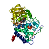
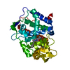
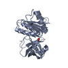
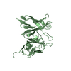
THE CONSTRUCT (RESIDUES 30-382) WAS EXPRESSED WITH AN N-TERMINAL PURIFICATION TAG ...THE CONSTRUCT (RESIDUES 30-382) WAS EXPRESSED WITH AN N-TERMINAL PURIFICATION TAG MGSDKIHHHHHHENLYFQG. THE TAG WAS REMOVED WITH TEV PROTEASE LEAVING ONLY A GLYCINE (0) FOLLOWED BY THE TARGET SEQUENCE.
-
Experimental details
-
Experiment
Experiment
Method: X-RAY DIFFRACTION / Number of used crystals: 1
-
Sample preparation
Crystal
Density Matthews: 3.59 Å3/Da / Density % sol: 65.78 %
Crystal grow
Temperature: 277 K / Method: vapor diffusion, sitting drop / pH: 6.3 Details: 20.0% polyethylene glycol 3350, 0.2M ammonium nitrate, No Buffer pH 6.3, NANODROP, VAPOR DIFFUSION, SITTING DROP, temperature 277K
Type: MARMOSAIC 325 mm CCD / Detector: CCD / Date: Jul 7, 2009 Details: Flat mirror (vertical focusing); single crystal Si(111) bent monochromator (ho rizontal focusing)
Radiation
Monochromator: single crystal Si(111) bent / Protocol: SINGLE WAVELENGTH / Monochromatic (M) / Laue (L): M / Scattering type: x-ray
Radiation wavelength
Wavelength: 0.97874 Å / Relative weight: 1
Reflection
Resolution: 2.46→29.656 Å / Num. obs: 40478 / % possible obs: 99.1 % / Redundancy: 4.2 % / Rmerge(I) obs: 0.094 / Rsym value: 0.094 / Net I/σ(I): 12.1
Reflection shell
Diffraction-ID: 1
Resolution (Å)
Redundancy (%)
Rmerge(I) obs
Mean I/σ(I) obs
Num. measured all
Num. unique all
Rsym value
% possible all
2.46-2.52
4.2
0.811
0.9
12540
2977
0.811
98.7
2.52-2.59
4.2
0.664
1.2
12105
2873
0.664
98.9
2.59-2.67
4.2
0.562
1.3
11943
2835
0.562
98.8
2.67-2.75
4.2
0.427
1.8
11476
2726
0.427
98.8
2.75-2.84
4.2
0.374
2
11078
2635
0.374
98.8
2.84-2.94
4.2
0.289
2.6
10911
2593
0.289
98.9
2.94-3.05
4.2
0.218
3.5
10404
2467
0.218
98.8
3.05-3.18
4.2
0.162
4.7
10063
2401
0.162
99.4
3.18-3.32
4.2
0.126
6
9663
2294
0.126
99.3
3.32-3.48
4.2
0.095
7.7
9159
2185
0.095
99.4
3.48-3.67
4.2
0.081
8.9
8948
2130
0.081
99.1
3.67-3.89
4.2
0.069
6.8
8179
1957
0.069
99.6
3.89-4.16
4.2
0.059
10.5
7835
1874
0.059
99.4
4.16-4.49
4.2
0.049
13.1
7282
1746
0.049
99.7
4.49-4.92
4.2
0.045
14.4
6744
1622
0.045
99.7
4.92-5.5
4.1
0.048
13.8
6023
1453
0.048
99.8
5.5-6.35
4.1
0.056
12
5375
1298
0.056
99.9
6.35-7.78
4.1
0.052
12.9
4495
1096
0.052
100
7.78-11
4
0.047
13
3440
854
0.047
99.9
11-29.661
3.8
0.043
14
1748
462
0.043
94.7
-
Phasing
Phasing
Method: SAD
-
Processing
Software
Name
Version
Classification
NB
MolProbity
3beta29
modelbuilding
PDB_EXTRACT
3.1
dataextraction
SHELX
phasing
SHARP
phasing
SCALA
3.2.5
datascaling
REFMAC
5.5.0110
refinement
MOSFLM
datareduction
SHELXD
phasing
Refinement
Method to determine structure: SAD / Resolution: 2.46→29.656 Å / Cor.coef. Fo:Fc: 0.913 / Cor.coef. Fo:Fc free: 0.903 / Occupancy max: 1 / Occupancy min: 0.5 / SU B: 13.866 / SU ML: 0.173 / Cross valid method: THROUGHOUT / σ(F): 0 / ESU R Free: 0.207 Stereochemistry target values: MAXIMUM LIKELIHOOD WITH PHASES Details: HYDROGENS HAVE BEEN ADDED IN THE RIDING POSITIONS U VALUES : WITH TLS ADDED OTHER REFINEMENT REMARKS: 1. HYDROGENS HAVE BEEN ADDED IN THE RIDING POSITIONS. 2. A MET-INHIBITION PROTOCOL WAS ...Details: HYDROGENS HAVE BEEN ADDED IN THE RIDING POSITIONS U VALUES : WITH TLS ADDED OTHER REFINEMENT REMARKS: 1. HYDROGENS HAVE BEEN ADDED IN THE RIDING POSITIONS. 2. A MET-INHIBITION PROTOCOL WAS USED FOR SELENOMETHIONINE INCORPORATION DURING PROTEIN EXPRESSION. THE OCCUPANCY OF THE SE ATOMS IN THE MSE RESIDUES WAS REDUCED TO 0.75 FOR THE REDUCED SCATTERING POWER DUE TO PARTIAL S-MET INCORPORATION. 3. SODIUM IONS MODELED ARE PRESENT IN PROTEIN BUFFER. 4. ATOM RECORD CONTAINS SUM OF TLS AND RESIDUAL B FACTORS. ANISOU RECORD CONTAINS SUM OF TLS AND RESIDUAL U FACTORS. 5. RESIDUES (A0-A124 AND B0-B122) WERE DISORDERED AND NOT INCLUDED IN THE FINAL MODEL. 6. TLS GROUPS WERE ASSIGNED WITH THE AID OF THE TLSMD SERVER.
Rfactor
Num. reflection
% reflection
Selection details
Rfree
0.2506
2033
5 %
RANDOM
Rwork
0.2325
-
-
-
obs
0.2334
40475
99.21 %
-
Solvent computation
Ion probe radii: 0.8 Å / Shrinkage radii: 0.8 Å / VDW probe radii: 1.4 Å / Solvent model: BABINET MODEL WITH MASK
In the structure databanks used in Yorodumi, some data are registered as the other names, "COVID-19 virus" and "2019-nCoV". Here are the details of the virus and the list of structure data.
Jan 31, 2019. EMDB accession codes are about to change! (news from PDBe EMDB page)
EMDB accession codes are about to change! (news from PDBe EMDB page)
The allocation of 4 digits for EMDB accession codes will soon come to an end. Whilst these codes will remain in use, new EMDB accession codes will include an additional digit and will expand incrementally as the available range of codes is exhausted. The current 4-digit format prefixed with “EMD-” (i.e. EMD-XXXX) will advance to a 5-digit format (i.e. EMD-XXXXX), and so on. It is currently estimated that the 4-digit codes will be depleted around Spring 2019, at which point the 5-digit format will come into force.
The EM Navigator/Yorodumi systems omit the EMD- prefix.
Related info.:Q: What is EMD? / ID/Accession-code notation in Yorodumi/EM Navigator
Yorodumi is a browser for structure data from EMDB, PDB, SASBDB, etc.
This page is also the successor to EM Navigator detail page, and also detail information page/front-end page for Omokage search.
The word "yorodu" (or yorozu) is an old Japanese word meaning "ten thousand". "mi" (miru) is to see.
Related info.:EMDB / PDB / SASBDB / Comparison of 3 databanks / Yorodumi Search / Aug 31, 2016. New EM Navigator & Yorodumi / Yorodumi Papers / Jmol/JSmol / Function and homology information / Changes in new EM Navigator and Yorodumi
 Movie
Movie Controller

 Yorodumi
 Open data
 Basic information
 Components
 Keywords
 Function and homology information
 Bacteroides vulgatus (bacteria)
 X-RAY DIFFRACTION /
  Authors
 Citation
 Structure visualization
 Downloads & links
 Other downloads

 PDBj
PDBj Assembly


 Mass: 22.990 Da / Num. of mol.: 2 / Source method: obtained synthetically / Formula: Na
 Mass: 18.015 Da / Num. of mol.: 168 / Source method: isolated from a natural source / Formula: H2O
 Sample preparation
 / Beamline: BL11-1 / Wavelength: 0.97874
  Processing