 Movie
Movie Controller

[English] 日本語
 Yorodumi
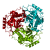
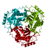
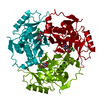
- PDB-1qca: QUADRUPLE MUTANT Q92C, N146F, Y168F, I172V TYPE III CAT COMPLEXED... -
+ Open data

- Basic information
| Entry | Database: PDB / ID: 1qca | ||||||
|---|---|---|---|---|---|---|---|
| Title | QUADRUPLE MUTANT Q92C, N146F, Y168F, I172V TYPE III CAT COMPLEXED WITH FUSIDIC ACID. CRYSTALS GROWN AT PH 6.3. X-RAY DATA COLLECTED AT ROOM TEMPERATURE | ||||||
 Components Components | TYPE III CHLORAMPHENICOL ACETYLTRANSFERASE | ||||||
 Keywords Keywords | TRANSFERASE (ACYLTRANSFERASE) / CHLORAMPHENICOL / FUSIDATE / STEROID | ||||||
| Function / homology |  Function and homology information Function and homology informationchloramphenicol O-acetyltransferase / chloramphenicol O-acetyltransferase activity / response to antibiotic Similarity search - Function | ||||||
| Biological species |  Shigella flexneri (bacteria) Shigella flexneri (bacteria) | ||||||
| Method |  X-RAY DIFFRACTION / Resolution: 2.2 Å X-RAY DIFFRACTION / Resolution: 2.2 Å | ||||||
 Authors Authors | Leslie, A.G.W. | ||||||
 Citation Citation | Journal: J.Mol.Biol. / Year: 1995 Title: Steroid recognition by chloramphenicol acetyltransferase: engineering and structural analysis of a high affinity fusidic acid binding site. Authors: Murray, I.A. / Cann, P.A. / Day, P.J. / Derrick, J.P. / Sutcliffe, M.J. / Shaw, W.V. / Leslie, A.G. #1: Journal: Annu.Rev.Biophys.Biophys.Chem. / Year: 1991Title: Chloramphenicol Acetyltransferase Authors: Shaw, W.V. / Leslie, A.G.W. #2: Journal: J.Mol.Biol. / Year: 1990Title: Refined Crystal Structure of Type III Chloramphenicol Acetyltransferase at 1.75-Angstroms Resolution Authors: Leslie, A.G.W. #3: Journal: Proc.Natl.Acad.Sci.USA / Year: 1988Title: Structure of Chloramphenicol Acetyltransferase at 1.75-Angstroms Resolution Authors: Leslie, A.G.W. / Moody, P.C.E. / Shaw, W.V. | ||||||
| History |
|
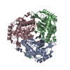
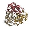
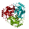
- Structure visualization
| Structure viewer | Molecule: MolmilJmol/JSmol |
|---|
- Downloads & links
-Download
| PDBx/mmCIF format | 1qca.cif.gz | 62.8 KB | Display | PDBx/mmCIF format |
|---|---|---|---|---|
| PDB format | pdb1qca.ent.gz | 44.4 KB | Display | PDB format |
| PDBx/mmJSON format | 1qca.json.gz | Tree view | PDBx/mmJSON format | |
| Others |  Other downloads Other downloads |
-Validation report
| Arichive directory | https://data.pdbj.org/pub/pdb/validation_reports/qc/1qcaftp://data.pdbj.org/pub/pdb/validation_reports/qc/1qca | HTTPS FTP |
|---|
-Related structure data
| Similar structure data |
|---|


-Links
 PDBj
PDBj


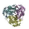
- Assembly
| Deposited unit | 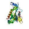
| |||||||||||||||||||||
|---|---|---|---|---|---|---|---|---|---|---|---|---|---|---|---|---|---|---|---|---|---|---|
| 1 | 
| |||||||||||||||||||||
| 2 | x 6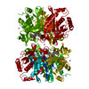
| |||||||||||||||||||||
| Unit cell |
| |||||||||||||||||||||
| Components on special symmetry positions |
| |||||||||||||||||||||
| Details | SYMMETRY THE CRYSTALLOGRAPHIC SYMMETRY TRANSFORMATIONS PRESENTED BELOW GENERATE THE SUBUNITS OF THE POLYMERIC MOLECULE. APPLIED TO RESIDUES: MET 6 .. LYS 219 120 DEGREE ROTATION ABOUT Z SYMMETRY1 1 -0.499953 -0.866025 0.000000 0.00000 SYMMETRY2 1 0.866026 -0.500047 0.000000 0.00000 SYMMETRY3 1 0.000000 0.000000 1.000000 0.00000 APPLIED TO RESIDUES: MET 6 .. LYS 219 120 DEGREE ROTATION ABOUT Z SYMMETRY1 2 -0.500047 0.866025 0.000000 0.00000 SYMMETRY2 2 -0.866026 -0.499953 0.000000 0.00000 SYMMETRY3 2 0.000000 0.000000 1.000000 0.00000 |
-Components
| #1: Protein | Mass: 24999.553 Da / Num. of mol.: 1 / Mutation: Q92C, N146F, Y168F, I172V Source method: isolated from a genetically manipulated source Source: (gene. exp.) Shigella flexneri (bacteria)Description: TRANSMISSIBLE PLASMID R387 ISOLATED FROM SHIGELLA FLEXNERI Plasmid: PUC18 / Production host: References: UniProt: P00484, chloramphenicol O-acetyltransferase | ||||||||
|---|---|---|---|---|---|---|---|---|---|
| #2: Chemical |   Mass: 58.933 Da / Num. of mol.: 2 / Source method: obtained synthetically / Formula: Co Mass: 58.933 Da / Num. of mol.: 2 / Source method: obtained synthetically / Formula: Co#3: Chemical | ChemComp-FUA / | 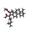  Mass: 516.709 Da / Num. of mol.: 1 / Source method: obtained synthetically / Formula: C31H48O6 / Comment: antibiotic, Antimicrobial*YM Mass: 516.709 Da / Num. of mol.: 1 / Source method: obtained synthetically / Formula: C31H48O6 / Comment: antibiotic, Antimicrobial*YM#4: Water | ChemComp-HOH / |  Mass: 18.015 Da / Num. of mol.: 144 / Source method: isolated from a natural source / Formula: H2O Mass: 18.015 Da / Num. of mol.: 144 / Source method: isolated from a natural source / Formula: H2ONonpolymer details | THERE ARE TWO COBALT IONS (BOTH OF WHICH LIE ON CRYSTALLOGRAPHIC TWO-FOLD AXES) WHICH PLAY A ...THERE ARE TWO COBALT IONS (BOTH OF WHICH LIE ON CRYSTALLOG | Sequence details | THE AMINO ACID NUMBERING SCHEME ADOPTED IS BASED ON THE ALIGNMENT OF SEVERAL CAT SEQUENCES. FOR THE ...THE AMINO ACID NUMBERING SCHEME ADOPTED IS BASED ON THE ALIGNMENT OF SEVERAL CAT SEQUENCES. FOR THE TYPE III ENZYME WHOSE COORDINATE | |
-Experimental details
-Experiment
| Experiment | Method: X-RAY DIFFRACTION / Number of used crystals: 1 |
|---|
- Sample preparation
| Crystal | Density Matthews: 2.78 Å3/Da / Density % sol: 55.69 % | ||||||||||||||||||||||||||||||||||||||||||||||||
|---|---|---|---|---|---|---|---|---|---|---|---|---|---|---|---|---|---|---|---|---|---|---|---|---|---|---|---|---|---|---|---|---|---|---|---|---|---|---|---|---|---|---|---|---|---|---|---|---|---|
| Crystal grow | pH: 6.3 / Details: pH 6.3 | ||||||||||||||||||||||||||||||||||||||||||||||||
| Crystal grow | *PLUS Temperature: 4 ℃ / Method: microdialysis | ||||||||||||||||||||||||||||||||||||||||||||||||
| Components of the solutions | *PLUS
|
-Data collection
| Diffraction | Mean temperature: 298 K |
|---|---|
| Diffraction source | Wavelength: 1.5418 Å |
| Detector | Detector: IMAGE PLATE / Date: Aug 27, 1992 |
| Radiation | Monochromatic (M) / Laue (L): M / Scattering type: x-ray |
| Radiation wavelength | Wavelength: 1.5418 Å / Relative weight: 1 |
| Reflection | Resolution: 2.2→15.6 Å / Num. obs: 14238 / % possible obs: 99 % / Observed criterion σ(I): 0 / Redundancy: 4.8 % / Rmerge(I) obs: 0.095 |
| Reflection | *PLUS Num. measured all: 68511 |
- Processing
| Software |
| ||||||||||||||||||||||||||||||||||||||||||||||||||||||||||||||||||||||||||||||||||||
|---|---|---|---|---|---|---|---|---|---|---|---|---|---|---|---|---|---|---|---|---|---|---|---|---|---|---|---|---|---|---|---|---|---|---|---|---|---|---|---|---|---|---|---|---|---|---|---|---|---|---|---|---|---|---|---|---|---|---|---|---|---|---|---|---|---|---|---|---|---|---|---|---|---|---|---|---|---|---|---|---|---|---|---|---|---|
| Refinement | Resolution: 2.2→6 Å / σ(F): 0 Details: THE STRUCTURE WAS SOLVED BY MOLECULAR REPLACEMENT USING THE REFINED 1.75 ANGSTROMS RESOLUTION STRUCTURE OF THE WILD-TYPE ENZYME AS A STARTING MODEL. CRYST1 TEXT TO EXPLAIN UNUSUAL UNIT-CELL ...Details: THE STRUCTURE WAS SOLVED BY MOLECULAR REPLACEMENT USING THE REFINED 1.75 ANGSTROMS RESOLUTION STRUCTURE OF THE WILD-TYPE ENZYME AS A STARTING MODEL. CRYST1 TEXT TO EXPLAIN UNUSUAL UNIT-CELL DATA: HEXAGONAL SETTING FOR R32 HOH 328 LIES ON A CRYSTALLOGRAPHIC TWO-FOLD AXIS AND IS NOT A TRUE SOLVENT MOLECULE, BUT IS IN MODELLING DENSITY SURROUNDING THE COBALT ION CO223 (ALSO ON THE TWO-FOLD AXIS). PRESUMABLY THIS DENSITY IS DUE TO THE HEXAMMINE GROUP (THE CRYSTALS WERE GROWN IN THE PRESENCE OF COBALT HEXAMMINE).
| ||||||||||||||||||||||||||||||||||||||||||||||||||||||||||||||||||||||||||||||||||||
| Displacement parameters | Biso mean: 19.9 Å2 | ||||||||||||||||||||||||||||||||||||||||||||||||||||||||||||||||||||||||||||||||||||
| Refinement step | Cycle: LAST / Resolution: 2.2→6 Å
| ||||||||||||||||||||||||||||||||||||||||||||||||||||||||||||||||||||||||||||||||||||
| Refine LS restraints |
| ||||||||||||||||||||||||||||||||||||||||||||||||||||||||||||||||||||||||||||||||||||
| Software | *PLUS Name: PROTIN/PROLSQ / Classification: refinement | ||||||||||||||||||||||||||||||||||||||||||||||||||||||||||||||||||||||||||||||||||||
| Refine LS restraints | *PLUS Type: p_plane_restr / Dev ideal target: 0.02 |
