 Movie
Movie Controller

[English] 日本語
 Yorodumi
- PDB-1pd5: Crystal structure of E.coli chloramphenicol acetyltransferase typ... -
+ Open data

- Basic information
| Entry | Database: PDB / ID: 1pd5 | ||||||
|---|---|---|---|---|---|---|---|
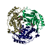
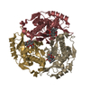
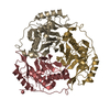
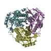
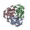
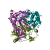
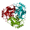
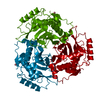
| Title | Crystal structure of E.coli chloramphenicol acetyltransferase type I at 2.5 Angstrom resolution | ||||||
 Components Components | Chloramphenicol acetyltransferase | ||||||
 Keywords Keywords | TRANSFERASE / trimer / chloramphenicol / acetyltransferase | ||||||
| Function / homology |  Function and homology information Function and homology informationchloramphenicol O-acetyltransferase / chloramphenicol O-acetyltransferase activity / response to antibiotic Similarity search - Function | ||||||
| Biological species |  | ||||||
| Method |  X-RAY DIFFRACTION / SYNCHROTRON / MOLECULAR REPLACEMENT / Resolution: 2.5 Å X-RAY DIFFRACTION / SYNCHROTRON / MOLECULAR REPLACEMENT / Resolution: 2.5 Å | ||||||
 Authors Authors | Roidis, A. / Kokkinidis, M. | ||||||
 Citation Citation | Journal: To be Published Title: Crystal structure of E.coli chloramphenicol acetyltransferase type I at 2.5 Angstrom resolution Authors: Roidis, A. / Kokkinidis, M. #1: Journal: Acta Crystallogr.,Sect.D / Year: 2000Title: Crystallization of type I chloramphenicol acetyltransferase:an approach based on the concept of ionic strength reducers Authors: Andreeva, A.E. / Borissova, B.E. / Mironova, R. / Glykos, N.M. / Kotsifaki, D. / Ivanov, I. / Krysteva, M. / Kokkinidis, M. | ||||||
| History |
|
- Structure visualization
| Structure viewer | Molecule: MolmilJmol/JSmol |
|---|
- Downloads & links
-Download
| PDBx/mmCIF format | 1pd5.cif.gz | 518.5 KB | Display | PDBx/mmCIF format |
|---|---|---|---|---|
| PDB format | pdb1pd5.ent.gz | 428.2 KB | Display | PDB format |
| PDBx/mmJSON format | 1pd5.json.gz | Tree view | PDBx/mmJSON format | |
| Others |  Other downloads Other downloads |
-Validation report
| Arichive directory | https://data.pdbj.org/pub/pdb/validation_reports/pd/1pd5ftp://data.pdbj.org/pub/pdb/validation_reports/pd/1pd5 | HTTPS FTP |
|---|
-Related structure data
| Related structure data |  3claS S: Starting model for refinement |
|---|---|
| Similar structure data |


-Links
 PDBj
PDBj

- Assembly
| Deposited unit | 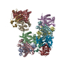
| ||||||||
|---|---|---|---|---|---|---|---|---|---|
| 1 | 
| ||||||||
| 2 | 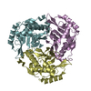
| ||||||||
| 3 | 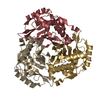
| ||||||||
| 4 | 
| ||||||||
| Unit cell |
| ||||||||
| Details | The biological assembly is a trimer. There are four trimers in the asymmetric unit ABC,DEF,GHI,JKL |
-Components
| #1: Protein | Mass: 25692.145 Da / Num. of mol.: 12 Source method: isolated from a genetically manipulated source Source: (gene. exp.) References: UniProt: P62577, chloramphenicol O-acetyltransferase #2: Water | ChemComp-HOH / |  Mass: 18.015 Da / Num. of mol.: 208 / Source method: isolated from a natural source / Formula: H2O Mass: 18.015 Da / Num. of mol.: 208 / Source method: isolated from a natural source / Formula: H2O |
|---|
-Experimental details
-Experiment
| Experiment | Method: X-RAY DIFFRACTION / Number of used crystals: 1 |
|---|
- Sample preparation
| Crystal | Density Matthews: 2.73 Å3/Da / Density % sol: 54.86 % |
|---|---|
| Crystal grow | Temperature: 290 K / Method: vapor diffusion, hanging drop / pH: 5.8 Details: methanol,calcium chloride,MES, pH 5.8, VAPOR DIFFUSION, HANGING DROP, temperature 290.0K |
-Data collection
| Diffraction | Mean temperature: 100 K |
|---|---|
| Diffraction source | Source: SYNCHROTRON / Site: EMBL/DESY, HAMBURG  / Beamline: X11 / Wavelength: 0.81 Å / Beamline: X11 / Wavelength: 0.81 Å |
| Detector | Type: MARRESEARCH / Detector: CCD / Date: Sep 28, 2002 / Details: Bent mirror |
| Radiation | Monochromator: Triangular monochromator / Protocol: SINGLE WAVELENGTH / Monochromatic (M) / Laue (L): M / Scattering type: x-ray |
| Radiation wavelength | Wavelength: 0.81 Å / Relative weight: 1 |
| Reflection | Resolution: 2.5→10 Å / Num. all: 114277 / Num. obs: 114277 / % possible obs: 99.6 % / Observed criterion σ(F): 13.91 / Observed criterion σ(I): 12.6 / Redundancy: 3.28 % / Rmerge(I) obs: 0.065 / Rsym value: 0.061 |
| Reflection shell | Resolution: 2.5→2.59 Å / Rmerge(I) obs: 0.291 / Num. unique all: 9674 / Rsym value: 0.246 / % possible all: 99.2 |
- Processing
| Software |
| ||||||||||||||||||||||||||||||||||||||||||||||||||||||||||||||||||||||
|---|---|---|---|---|---|---|---|---|---|---|---|---|---|---|---|---|---|---|---|---|---|---|---|---|---|---|---|---|---|---|---|---|---|---|---|---|---|---|---|---|---|---|---|---|---|---|---|---|---|---|---|---|---|---|---|---|---|---|---|---|---|---|---|---|---|---|---|---|---|---|---|
| Refinement | Method to determine structure: MOLECULAR REPLACEMENT Starting model: PDB ENTRY 3CLA Resolution: 2.5→10 Å / Cor.coef. Fo:Fc: 0.947 / Cor.coef. Fo:Fc free: 0.892 / SU B: 9.945 / SU ML: 0.224 / Isotropic thermal model: ISOTROPIC / Cross valid method: THROUGHOUT / σ(F): 2 / ESU R: 0.518 / ESU R Free: 0.324 / Stereochemistry target values: MAXIMUM LIKELIHOOD
| ||||||||||||||||||||||||||||||||||||||||||||||||||||||||||||||||||||||
| Solvent computation | Ion probe radii: 0.8 Å / Shrinkage radii: 0.8 Å / VDW probe radii: 1.4 Å / Solvent model: BABINET MODEL WITH MASK | ||||||||||||||||||||||||||||||||||||||||||||||||||||||||||||||||||||||
| Displacement parameters | Biso mean: 46.425 Å2
| ||||||||||||||||||||||||||||||||||||||||||||||||||||||||||||||||||||||
| Refinement step | Cycle: LAST / Resolution: 2.5→10 Å
| ||||||||||||||||||||||||||||||||||||||||||||||||||||||||||||||||||||||
| Refine LS restraints |
| ||||||||||||||||||||||||||||||||||||||||||||||||||||||||||||||||||||||
| LS refinement shell | Resolution: 2.5→2.561 Å / Total num. of bins used: 20 /
|
