 Movie
Movie Controller

[English] 日本語
 Yorodumi
- PDB-4wy7: Crystal structure of recombinant 4E10 expressed in Escherichia co... -
+ Open data

- Basic information
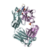
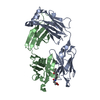
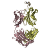
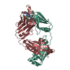
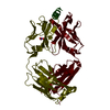
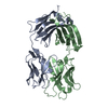
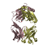
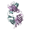
| Entry | Database: PDB / ID: 4wy7 | ||||||
|---|---|---|---|---|---|---|---|
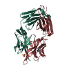
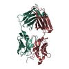
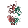
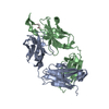
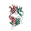
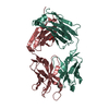
| Title | Crystal structure of recombinant 4E10 expressed in Escherichia coli with epitope bound | ||||||
 Components Components |
| ||||||
 Keywords Keywords | IMMUNE SYSTEM / broad neutralizing antobody / recombinant Fab / epitope | ||||||
| Function / homology |  Function and homology information Function and homology informationDectin-2 family / positive regulation of plasma membrane raft polarization / symbiont-mediated perturbation of host defense response / positive regulation of receptor clustering / host cell endosome membrane / clathrin-dependent endocytosis of virus by host cell / viral protein processing / fusion of virus membrane with host plasma membrane / fusion of virus membrane with host endosome membrane / viral envelope ...Dectin-2 family / positive regulation of plasma membrane raft polarization / symbiont-mediated perturbation of host defense response / positive regulation of receptor clustering / host cell endosome membrane / clathrin-dependent endocytosis of virus by host cell / viral protein processing / fusion of virus membrane with host plasma membrane / fusion of virus membrane with host endosome membrane / viral envelope / virion attachment to host cell / host cell plasma membrane / virion membrane / structural molecule activity / membrane Similarity search - Function | ||||||
| Biological species |  Homo sapiens (human) Homo sapiens (human) Human immunodeficiency virus type 1 group M subtype B Human immunodeficiency virus type 1 group M subtype B | ||||||
| Method |  X-RAY DIFFRACTION / SYNCHROTRON / MOLECULAR REPLACEMENT / Resolution: 2.1 Å X-RAY DIFFRACTION / SYNCHROTRON / MOLECULAR REPLACEMENT / Resolution: 2.1 Å | ||||||
 Authors Authors | Rujas, E. / Morante, K. / Tsumoto, K. / Nieva, J.L. / Caaveiro, J.M.M. | ||||||
 Citation Citation | Journal: J.Biol.Chem. / Year: 2015 Title: The Atomic Structure of the HIV-1 gp41 Transmembrane Domain and Its Connection to the Immunogenic Membrane-proximal External Region. Authors: Apellaniz, B. / Rujas, E. / Serrano, S. / Morante, K. / Tsumoto, K. / Caaveiro, J.M. / Jimenez, M.A. / Nieva, J.L. | ||||||
| History |
|
- Structure visualization
| Structure viewer | Molecule: MolmilJmol/JSmol |
|---|
- Downloads & links
-Download
| PDBx/mmCIF format | 4wy7.cif.gz | 108.7 KB | Display | PDBx/mmCIF format |
|---|---|---|---|---|
| PDB format | pdb4wy7.ent.gz | 80.8 KB | Display | PDB format |
| PDBx/mmJSON format | 4wy7.json.gz | Tree view | PDBx/mmJSON format | |
| Others |  Other downloads Other downloads |
-Validation report
| Arichive directory | https://data.pdbj.org/pub/pdb/validation_reports/wy/4wy7ftp://data.pdbj.org/pub/pdb/validation_reports/wy/4wy7 | HTTPS FTP |
|---|
-Related structure data
| Related structure data |  2mg1C  2mg2C  2mg3C 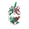 2fx7S S: Starting model for refinement C: citing same article ( |
|---|---|
| Similar structure data |


-Links
 PDBj
PDBj


- Assembly
| Deposited unit | 
| ||||||||
|---|---|---|---|---|---|---|---|---|---|
| 1 |
| ||||||||
| Unit cell |
|
-Components
| #1: Antibody | Mass: 23990.027 Da / Num. of mol.: 1 Source method: isolated from a genetically manipulated source Source: (gene. exp.) Homo sapiens (human) / Plasmid: pET-Duet1 / Production host:  | ||||
|---|---|---|---|---|---|
| #2: Antibody | Mass: 23292.705 Da / Num. of mol.: 1 Source method: isolated from a genetically manipulated source Source: (gene. exp.) Homo sapiens (human) / Plasmid: pET-Duet1 / Production host: | ||||
| #3: Protein/peptide | Mass: 2187.582 Da / Num. of mol.: 1 / Fragment: UNP residues 662-674 Source method: isolated from a genetically manipulated source Source: (gene. exp.) Human immunodeficiency virus type 1 group M subtype BStrain: isolate WMJ22 / Gene: env / Production host: synthetic construct (others) / References: UniProt: P05880 | ||||
| #4: Chemical |   Mass: 92.094 Da / Num. of mol.: 2 / Source method: obtained synthetically / Formula: C3H8O3 Mass: 92.094 Da / Num. of mol.: 2 / Source method: obtained synthetically / Formula: C3H8O3#5: Water | ChemComp-HOH / |  Mass: 18.015 Da / Num. of mol.: 304 / Source method: isolated from a natural source / Formula: H2O Mass: 18.015 Da / Num. of mol.: 304 / Source method: isolated from a natural source / Formula: H2OHas protein modification | Y | |
-Experimental details
-Experiment
| Experiment | Method: X-RAY DIFFRACTION |
|---|
- Sample preparation
| Crystal | Density Matthews: 2.77 Å3/Da / Density % sol: 55.58 % |
|---|---|
| Crystal grow | Temperature: 293.15 K / Method: vapor diffusion, hanging drop / pH: 6.5 Details: 200 mM sodium acetate 30% PEG 8,000 100 mM sodium cacodylate Protein was prepared in PBS |
-Data collection
| Diffraction | Mean temperature: 100 K |
|---|---|
| Diffraction source | Source: SYNCHROTRON / Site: Photon Factory  / Beamline: BL-5A / Wavelength: 1 Å / Beamline: BL-5A / Wavelength: 1 Å |
| Detector | Type: ADSC QUANTUM 315r / Detector: CCD / Date: Jun 28, 2014 |
| Radiation | Protocol: SINGLE WAVELENGTH / Monochromatic (M) / Laue (L): M / Scattering type: x-ray |
| Radiation wavelength | Wavelength: 1 Å / Relative weight: 1 |
| Reflection | Resolution: 2.1→44.7 Å / Num. obs: 30114 / % possible obs: 94.2 % / Redundancy: 5.5 % / Rmerge(I) obs: 0.102 / Net I/σ(I): 14 |
| Reflection shell | Resolution: 2.1→2.21 Å / Redundancy: 5.3 % / Rmerge(I) obs: 0.37 / Mean I/σ(I) obs: 4.3 / % possible all: 76.6 |
- Processing
| Software |
| ||||||||||||||||||||||||||||||||||||||||||||||||||||||||||||||||||||||||||||||||||||||||||||||||||||||||||||||||||||||||||||||||||||||||||||||||||||||||||||||||||||||||||||||||||||||
|---|---|---|---|---|---|---|---|---|---|---|---|---|---|---|---|---|---|---|---|---|---|---|---|---|---|---|---|---|---|---|---|---|---|---|---|---|---|---|---|---|---|---|---|---|---|---|---|---|---|---|---|---|---|---|---|---|---|---|---|---|---|---|---|---|---|---|---|---|---|---|---|---|---|---|---|---|---|---|---|---|---|---|---|---|---|---|---|---|---|---|---|---|---|---|---|---|---|---|---|---|---|---|---|---|---|---|---|---|---|---|---|---|---|---|---|---|---|---|---|---|---|---|---|---|---|---|---|---|---|---|---|---|---|---|---|---|---|---|---|---|---|---|---|---|---|---|---|---|---|---|---|---|---|---|---|---|---|---|---|---|---|---|---|---|---|---|---|---|---|---|---|---|---|---|---|---|---|---|---|---|---|---|---|
| Refinement | Method to determine structure: MOLECULAR REPLACEMENT Starting model: 2FX7 Resolution: 2.1→44.7 Å / Cor.coef. Fo:Fc: 0.955 / Cor.coef. Fo:Fc free: 0.932 / SU B: 4.171 / SU ML: 0.11 / Cross valid method: THROUGHOUT / ESU R: 0.187 / ESU R Free: 0.165 / Stereochemistry target values: MAXIMUM LIKELIHOOD / Details: HYDROGENS HAVE BEEN ADDED IN THE RIDING POSITIONS
| ||||||||||||||||||||||||||||||||||||||||||||||||||||||||||||||||||||||||||||||||||||||||||||||||||||||||||||||||||||||||||||||||||||||||||||||||||||||||||||||||||||||||||||||||||||||
| Solvent computation | Ion probe radii: 0.8 Å / Shrinkage radii: 0.8 Å / VDW probe radii: 1.2 Å / Solvent model: BABINET MODEL WITH MASK | ||||||||||||||||||||||||||||||||||||||||||||||||||||||||||||||||||||||||||||||||||||||||||||||||||||||||||||||||||||||||||||||||||||||||||||||||||||||||||||||||||||||||||||||||||||||
| Displacement parameters | Biso mean: 31.78 Å2
| ||||||||||||||||||||||||||||||||||||||||||||||||||||||||||||||||||||||||||||||||||||||||||||||||||||||||||||||||||||||||||||||||||||||||||||||||||||||||||||||||||||||||||||||||||||||
| Refinement step | Cycle: 1 / Resolution: 2.1→44.7 Å
| ||||||||||||||||||||||||||||||||||||||||||||||||||||||||||||||||||||||||||||||||||||||||||||||||||||||||||||||||||||||||||||||||||||||||||||||||||||||||||||||||||||||||||||||||||||||
| Refine LS restraints |
|