 ムービー
ムービー コントローラー


+ データを開く

- 基本情報
基本情報
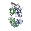
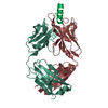
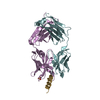
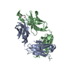
| 登録情報 | データベース: PDB / ID: 2fx7 | ||||||
|---|---|---|---|---|---|---|---|
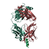
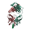
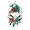
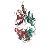
| タイトル | Crystal structure of hiv-1 neutralizing human fab 4e10 in complex with a 16-residue peptide encompassing the 4e10 epitope on gp41 | ||||||
 要素 要素 |
| ||||||
 キーワード キーワード | IMMUNE SYSTEM / immunoglobulin fold / beta-sandwich / antibody-epitope complex | ||||||
| 機能・相同性 |  機能・相同性情報 機能・相同性情報Dectin-2 family / symbiont-mediated perturbation of host defense response / positive regulation of plasma membrane raft polarization / positive regulation of receptor clustering / host cell endosome membrane / clathrin-dependent endocytosis of virus by host cell / viral protein processing / fusion of virus membrane with host plasma membrane / fusion of virus membrane with host endosome membrane / viral envelope ...Dectin-2 family / symbiont-mediated perturbation of host defense response / positive regulation of plasma membrane raft polarization / positive regulation of receptor clustering / host cell endosome membrane / clathrin-dependent endocytosis of virus by host cell / viral protein processing / fusion of virus membrane with host plasma membrane / fusion of virus membrane with host endosome membrane / viral envelope / virion attachment to host cell / host cell plasma membrane / virion membrane / structural molecule activity / membrane 類似検索 - 分子機能 | ||||||
| 生物種 |  Homo sapiens (ヒト) Homo sapiens (ヒト) | ||||||
| 手法 |  X線回折 / シンクロトロン / 分子置換 / 解像度: 1.76 Å X線回折 / シンクロトロン / 分子置換 / 解像度: 1.76 Å | ||||||
 データ登録者 データ登録者 | Cardoso, R.M.F. / Brunel, F.M. / Ferguson, S. / Burton, D.R. / Dawson, P.E. / Wilson, I.A. | ||||||
 引用 引用 | ジャーナル: J.Mol.Biol. / 年: 2007 タイトル: Structural basis of enhanced binding of extended and helically constrained peptide epitopes of the broadly neutralizing HIV-1 antibody 4E10. 著者: Cardoso, R.M. / Brunel, F.M. / Ferguson, S. / Zwick, M. / Burton, D.R. / Dawson, P.E. / Wilson, I.A. #1: ジャーナル: Immunity / 年: 2005タイトル: Broadly neutralizing anti-HIV antibody 4E10 recognizes a helical conformation of a highly conserved fusion-associated motif in gp41 著者: Cardoso, R.M.F. / Zwick, M.B. / Stanfield, R.L. / Kunert, R. / Binley, J.M. / Katinger, H. / Burton, D.R. / Wilson, I.A. | ||||||
| 履歴 |
| ||||||
| Remark 999 | No database reference is currently available for the light and heavy chains (Chain L and H). |
- 構造の表示
構造の表示
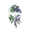
| 構造ビューア | 分子: MolmilJmol/JSmol |
|---|
- ダウンロードとリンク
-ダウンロード
| PDBx/mmCIF形式 | 2fx7.cif.gz | 108.3 KB | 表示 | PDBx/mmCIF形式 |
|---|---|---|---|---|
| PDB形式 | pdb2fx7.ent.gz | 82 KB | 表示 | PDB形式 |
| PDBx/mmJSON形式 | 2fx7.json.gz | ツリー表示 | PDBx/mmJSON形式 | |
| その他 |  その他のダウンロード その他のダウンロード |
-検証レポート
| アーカイブディレクトリ | https://data.pdbj.org/pub/pdb/validation_reports/fx/2fx7ftp://data.pdbj.org/pub/pdb/validation_reports/fx/2fx7 | HTTPS FTP |
|---|
-関連構造データ


-リンク
 PDBj
PDBj


- 集合体
集合体
| 登録構造単位 | 
| ||||||||
|---|---|---|---|---|---|---|---|---|---|
| 1 |
| ||||||||
| 単位格子 |
|
-要素
| #1: 抗体 | 分子量: 23292.705 Da / 分子数: 1 / 断片: Light Chain / 由来タイプ: 組換発現 / 由来: (組換発現) Homo sapiens (ヒト)発現宿主:   Cricetulus griseus (モンゴルキヌゲネズミ) Cricetulus griseus (モンゴルキヌゲネズミ) |
|---|---|
| #2: 抗体 | 分子量: 23990.027 Da / 分子数: 1 / 断片: Heavy Chain / 由来タイプ: 組換発現 / 由来: (組換発現) Homo sapiens (ヒト)発現宿主: Cricetulus griseus (モンゴルキヌゲネズミ) |
| #3: タンパク質・ペプチド | 分子量: 2187.582 Da / 分子数: 1 / 断片: Peptide Epitope of 4E10 / Mutation: I684K, F685K, I686K / 由来タイプ: 合成 詳細: This sequence includes a fragment of the HIV envelope protein gp41 参照: UniProt: P05880 |
| #4: 化合物 | ChemComp-GOL /   分子量: 92.094 Da / 分子数: 1 / 由来タイプ: 合成 / 式: C3H8O3 分子量: 92.094 Da / 分子数: 1 / 由来タイプ: 合成 / 式: C3H8O3 |
| #5: 水 | ChemComp-HOH /  分子量: 18.015 Da / 分子数: 337 / 由来タイプ: 天然 / 式: H2O 分子量: 18.015 Da / 分子数: 337 / 由来タイプ: 天然 / 式: H2O |
| Has protein modification | Y |
-実験情報
-実験
| 実験 | 手法: X線回折 / 使用した結晶の数: 1 |
|---|
- 試料調製
試料調製
| 結晶 | マシュー密度: 2.79 Å3/Da / 溶媒含有率: 55.98 % |
|---|---|
| 結晶化 | 温度: 295 K / 手法: 蒸気拡散法, シッティングドロップ法 / pH: 5.6 詳細: 26% PEG 8000, 0.2 M sodium acetate, 0.2 M sodium thiocyanate, pH 5.6, VAPOR DIFFUSION, SITTING DROP, temperature 295K |
-データ収集
| 回折 | 平均測定温度: 100 K |
|---|---|
| 放射光源 | 由来: シンクロトロン / サイト: SSRL  / ビームライン: BL11-1 / 波長: 0.97945 Å / ビームライン: BL11-1 / 波長: 0.97945 Å |
| 検出器 | タイプ: ADSC QUANTUM 315 / 検出器: CCD / 日付: 2005年3月11日 |
| 放射 | モノクロメーター: Curved Crystal / プロトコル: SINGLE WAVELENGTH / 単色(M)・ラウエ(L): M / 散乱光タイプ: x-ray |
| 放射波長 | 波長: 0.97945 Å / 相対比: 1 |
| 反射 | 解像度: 1.76→50 Å / Num. all: 54368 / Num. obs: 53993 / % possible obs: 99.3 % / Observed criterion σ(F): 0 / Observed criterion σ(I): 3 / 冗長度: 3.7 % / Biso Wilson estimate: 18.1 Å2 / Rsym value: 0.065 / Net I/σ(I): 14.8 |
| 反射 シェル | 解像度: 1.76→1.82 Å / 冗長度: 3.7 % / Mean I/σ(I) obs: 5.2 / Num. unique all: 5390 / Rsym value: 0.302 / % possible all: 99.9 |
- 解析
解析
| ソフトウェア |
| |||||||||||||||||||||||||
|---|---|---|---|---|---|---|---|---|---|---|---|---|---|---|---|---|---|---|---|---|---|---|---|---|---|---|
| 精密化 | 構造決定の手法: 分子置換 開始モデル: PDB ENTRY 1TZG 解像度: 1.76→50 Å / Isotropic thermal model: Isotropic / 交差検証法: THROUGHOUT / σ(F): 0 / 立体化学のターゲット値: Engh & Huber
| |||||||||||||||||||||||||
| 溶媒の処理 | Bsol: 47.0027 Å2 / ksol: 0.398008 e/Å3 | |||||||||||||||||||||||||
| 原子変位パラメータ | Biso mean: 24.6 Å2
| |||||||||||||||||||||||||
| Refine analyze | Luzzati coordinate error obs: 0.202 Å | |||||||||||||||||||||||||
| 精密化ステップ | サイクル: LAST / 解像度: 1.76→50 Å
| |||||||||||||||||||||||||
| 拘束条件 |
| |||||||||||||||||||||||||
| LS精密化 シェル |
|
