 Movie
Movie Controller

[English] 日本語
 Yorodumi
- PDB-2fx9: Crystal structure of hiv-1 neutralizing human fab 4e10 in complex... -
+ Open data

- Basic information
| Entry | Database: PDB / ID: 2fx9 | ||||||
|---|---|---|---|---|---|---|---|
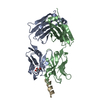
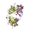
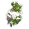
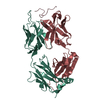
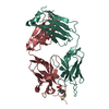
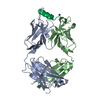
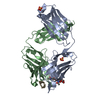
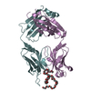
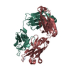
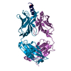
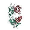
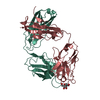
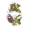
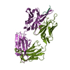
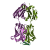
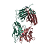
| Title | Crystal structure of hiv-1 neutralizing human fab 4e10 in complex with a thioether-linked peptide encompassing the 4e10 epitope on gp41 | ||||||
 Components Components |
| ||||||
 Keywords Keywords | IMMUNE SYSTEM / immunoglobulin fold / beta-sandwich / antibody-epitope complex | ||||||
| Function / homology |  Function and homology information Function and homology informationDectin-2 family / symbiont-mediated perturbation of host defense response / positive regulation of plasma membrane raft polarization / positive regulation of receptor clustering / host cell endosome membrane / clathrin-dependent endocytosis of virus by host cell / viral protein processing / fusion of virus membrane with host plasma membrane / fusion of virus membrane with host endosome membrane / viral envelope ...Dectin-2 family / symbiont-mediated perturbation of host defense response / positive regulation of plasma membrane raft polarization / positive regulation of receptor clustering / host cell endosome membrane / clathrin-dependent endocytosis of virus by host cell / viral protein processing / fusion of virus membrane with host plasma membrane / fusion of virus membrane with host endosome membrane / viral envelope / virion attachment to host cell / host cell plasma membrane / virion membrane / structural molecule activity / membrane Similarity search - Function | ||||||
| Biological species |  Homo sapiens (human) Homo sapiens (human) | ||||||
| Method |  X-RAY DIFFRACTION / SYNCHROTRON / MOLECULAR REPLACEMENT / Resolution: 2.1 Å X-RAY DIFFRACTION / SYNCHROTRON / MOLECULAR REPLACEMENT / Resolution: 2.1 Å | ||||||
 Authors Authors | Cardoso, R.M.F. / Brunel, F.M. / Ferguson, S. / Burton, D.R. / Dawson, P.E. / Wilson, I.A. | ||||||
 Citation Citation | Journal: J.Mol.Biol. / Year: 2007 Title: Structural basis of enhanced binding of extended and helically constrained peptide epitopes of the broadly neutralizing HIV-1 antibody 4E10. Authors: Cardoso, R.M. / Brunel, F.M. / Ferguson, S. / Zwick, M. / Burton, D.R. / Dawson, P.E. / Wilson, I.A. #1: Journal: Immunity / Year: 2005Title: Broadly neutralizing anti-HIV antibody 4E10 recognizes a helical conformation of a highly conserved fusion-associated motif in gp41 Authors: Cardoso, R.M.F. / Zwick, M.B. / Stanfield, R.L. / Kunert, R. / Binley, J.M. / Katinger, H. / Burton, D.R. / Wilson, I.A. | ||||||
| History |
| ||||||
| Remark 999 | No database reference is currently available for Chains L, M, H, I, P, and Q. |
- Structure visualization
| Structure viewer | Molecule: MolmilJmol/JSmol |
|---|
- Downloads & links
-Download
| PDBx/mmCIF format | 2fx9.cif.gz | 191.2 KB | Display | PDBx/mmCIF format |
|---|---|---|---|---|
| PDB format | pdb2fx9.ent.gz | 151.2 KB | Display | PDB format |
| PDBx/mmJSON format | 2fx9.json.gz | Tree view | PDBx/mmJSON format | |
| Others |  Other downloads Other downloads |
-Validation report
| Arichive directory | https://data.pdbj.org/pub/pdb/validation_reports/fx/2fx9ftp://data.pdbj.org/pub/pdb/validation_reports/fx/2fx9 | HTTPS FTP |
|---|
-Related structure data
| Related structure data | 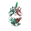 2fx7C 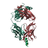 2fx8C 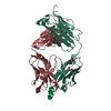 1tzgS S: Starting model for refinement C: citing same article ( |
|---|---|
| Similar structure data |


-Links
 PDBj
PDBj


- Assembly
| Deposited unit | 
| ||||||||
|---|---|---|---|---|---|---|---|---|---|
| 1 | 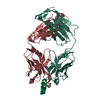
| ||||||||
| 2 | 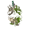
| ||||||||
| Unit cell |
|
-Components
| #1: Antibody | Mass: 23292.705 Da / Num. of mol.: 2 / Fragment: Light Chain Source method: isolated from a genetically manipulated source Source: (gene. exp.) Homo sapiens (human) / Production host:   Cricetulus griseus (Chinese hamster) Cricetulus griseus (Chinese hamster)#2: Antibody | Mass: 23860.848 Da / Num. of mol.: 2 / Fragment: Heavy Chain Source method: isolated from a genetically manipulated source Source: (gene. exp.) Homo sapiens (human) / Production host: Cricetulus griseus (Chinese hamster)#3: Protein/peptide | Mass: 1941.385 Da / Num. of mol.: 2 / Fragment: Peptide Epitope of 4E10 / Source method: obtained synthetically Details: This sequence includes a fragment of the HIV envelope protein gp41 References: UniProt: P05880*PLUS #4: Water | ChemComp-HOH / |  Mass: 18.015 Da / Num. of mol.: 403 / Source method: isolated from a natural source / Formula: H2O Mass: 18.015 Da / Num. of mol.: 403 / Source method: isolated from a natural source / Formula: H2O |
|---|
-Experimental details
-Experiment
| Experiment | Method: X-RAY DIFFRACTION / Number of used crystals: 1 |
|---|
- Sample preparation
| Crystal | Density Matthews: 2.31 Å3/Da / Density % sol: 46.71 % |
|---|---|
| Crystal grow | Temperature: 295 K / Method: vapor diffusion, sitting drop Details: 20% PEG 3350, 0.2 M ammonium nitrate, pH no buffer, VAPOR DIFFUSION, SITTING DROP, temperature 295K |
-Data collection
| Diffraction | Mean temperature: 100 K |
|---|---|
| Diffraction source | Source: SYNCHROTRON / Site: SSRL  / Beamline: BL9-2 / Wavelength: 0.95369 Å / Beamline: BL9-2 / Wavelength: 0.95369 Å |
| Detector | Type: MARMOSAIC 325 mm CCD / Detector: CCD / Date: May 5, 2005 |
| Radiation | Monochromator: double crystal / Protocol: SINGLE WAVELENGTH / Monochromatic (M) / Laue (L): M / Scattering type: x-ray |
| Radiation wavelength | Wavelength: 0.95369 Å / Relative weight: 1 |
| Reflection | Resolution: 2.1→50 Å / Num. all: 51978 / Num. obs: 51614 / % possible obs: 99.3 % / Observed criterion σ(F): 0 / Observed criterion σ(I): 3 / Redundancy: 3.7 % / Biso Wilson estimate: 31.6 Å2 / Rsym value: 0.094 / Net I/σ(I): 14.1 |
| Reflection shell | Resolution: 2.1→2.15 Å / Redundancy: 3.7 % / Mean I/σ(I) obs: 2.7 / Num. unique all: 3779 / Rsym value: 0.324 / % possible all: 99 |
- Processing
| Software |
| |||||||||||||||||||||||||
|---|---|---|---|---|---|---|---|---|---|---|---|---|---|---|---|---|---|---|---|---|---|---|---|---|---|---|
| Refinement | Method to determine structure: MOLECULAR REPLACEMENT Starting model: PBD ENTRY 1TZG Resolution: 2.1→50 Å / Isotropic thermal model: ISOTROPIC / Cross valid method: THROUGHOUT / σ(F): 0 / Stereochemistry target values: Engh & Huber
| |||||||||||||||||||||||||
| Solvent computation | Bsol: 46.1188 Å2 / ksol: 0.343825 e/Å3 | |||||||||||||||||||||||||
| Displacement parameters | Biso mean: 34.7 Å2
| |||||||||||||||||||||||||
| Refine analyze | Luzzati coordinate error obs: 0.316 Å | |||||||||||||||||||||||||
| Refinement step | Cycle: LAST / Resolution: 2.1→50 Å
| |||||||||||||||||||||||||
| Refine LS restraints |
| |||||||||||||||||||||||||
| LS refinement shell |
|
