 Movie
Movie Controller

[English] 日本語
 Yorodumi
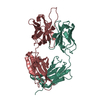
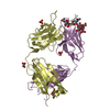
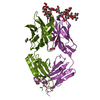
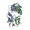
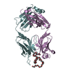
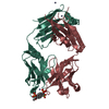
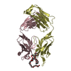
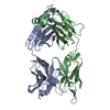
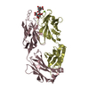
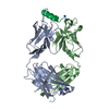
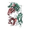
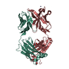
- PDB-1yeg: STRUCTURE OF IGG2A FAB FRAGMENT (D2.3) COMPLEXED WITH REACTION PRODUCT -
+ Open data

- Basic information
| Entry | Database: PDB / ID: 1yeg | ||||||
|---|---|---|---|---|---|---|---|
| Title | STRUCTURE OF IGG2A FAB FRAGMENT (D2.3) COMPLEXED WITH REACTION PRODUCT | ||||||
 Components Components | (IGG2A FAB FRAGMENT) x 2 | ||||||
 Keywords Keywords | CATALYTIC ANTIBODY | ||||||
| Function / homology |  Function and homology information Function and homology informationFc-gamma receptor I complex binding / immunoglobulin complex, circulating / immunoglobulin receptor binding / IgG immunoglobulin complex / complement activation, classical pathway / antigen binding / antibacterial humoral response / : Similarity search - Function | ||||||
| Biological species |  | ||||||
| Method |  X-RAY DIFFRACTION / SYNCHROTRON / MOLECULAR REPLACEMENT / Resolution: 2 Å X-RAY DIFFRACTION / SYNCHROTRON / MOLECULAR REPLACEMENT / Resolution: 2 Å | ||||||
 Authors Authors | Gigant, B. / Knossow, M. | ||||||
 Citation Citation | Journal: Proc.Natl.Acad.Sci.USA / Year: 1997 Title: X-ray structures of a hydrolytic antibody and of complexes elucidate catalytic pathway from substrate binding and transition state stabilization through water attack and product release. Authors: Gigant, B. / Charbonnier, J.B. / Eshhar, Z. / Green, B.S. / Knossow, M. | ||||||
| History |
|
- Structure visualization
| Structure viewer | Molecule: MolmilJmol/JSmol |
|---|
- Downloads & links
-Download
| PDBx/mmCIF format | 1yeg.cif.gz | 104.2 KB | Display | PDBx/mmCIF format |
|---|---|---|---|---|
| PDB format | pdb1yeg.ent.gz | 78.1 KB | Display | PDB format |
| PDBx/mmJSON format | 1yeg.json.gz | Tree view | PDBx/mmJSON format | |
| Others |  Other downloads Other downloads |
-Validation report
| Arichive directory | https://data.pdbj.org/pub/pdb/validation_reports/ye/1yegftp://data.pdbj.org/pub/pdb/validation_reports/ye/1yeg | HTTPS FTP |
|---|
-Related structure data
| Related structure data | 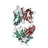 1yefC  1yehC 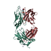 1yecS S: Starting model for refinement C: citing same article ( |
|---|---|
| Similar structure data |


-Links
 PDBj
PDBj


- Assembly
| Deposited unit | 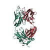
| ||||||||
|---|---|---|---|---|---|---|---|---|---|
| 1 |
| ||||||||
| 2 | 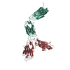
| ||||||||
| Unit cell |
|
-Components
-Antibody , 2 types, 2 molecules LH
| #1: Antibody | Mass: 24005.816 Da / Num. of mol.: 1 / Source method: isolated from a natural source / Details: STRUCTURE OF IGG2A FAB FRAGMENT (D2.3) / Source: (natural) |
|---|---|
| #2: Antibody | Mass: 24189.217 Da / Num. of mol.: 1 / Source method: isolated from a natural source / Details: STRUCTURE OF IGG2A FAB FRAGMENT (D2.3) / Source: (natural) |
-Non-polymers , 4 types, 163 molecules 


| #3: Chemical | ChemComp-ZN /  Mass: 65.409 Da / Num. of mol.: 7 / Source method: obtained synthetically / Formula: Zn Mass: 65.409 Da / Num. of mol.: 7 / Source method: obtained synthetically / Formula: Zn#4: Chemical | ChemComp-BPN / |  Mass: 153.135 Da / Num. of mol.: 1 / Source method: obtained synthetically / Formula: C7H7NO3 Mass: 153.135 Da / Num. of mol.: 1 / Source method: obtained synthetically / Formula: C7H7NO3#5: Chemical | ChemComp-ACT / |  Mass: 59.044 Da / Num. of mol.: 1 / Source method: obtained synthetically / Formula: C2H3O2 Mass: 59.044 Da / Num. of mol.: 1 / Source method: obtained synthetically / Formula: C2H3O2#6: Water | ChemComp-HOH / | Mass: 18.015 Da / Num. of mol.: 154 / Source method: isolated from a natural source / Formula: H2O |
|---|
-Details
| Has protein modification | Y |
|---|---|
| Sequence details | THE SEQUENCES OF THE CONSTANT DOMAINS OF THE HEAVY CHAINS. (RESIDUES H 106 - H 223) AND OF THE ...THE SEQUENCES OF THE CONSTANT DOMAINS OF THE HEAVY CHAINS. (RESIDUES H 106 - H 223) AND OF THE LIGHT CHAINS (RESIDUES L 107 - L 214) HAVE NOT BEEN DETERMINED |
-Experimental details
-Experiment
| Experiment | Method: X-RAY DIFFRACTION / Number of used crystals: 1 |
|---|
- Sample preparation
| Crystal | Density Matthews: 2.95 Å3/Da / Density % sol: 52.5 % | |||||||||||||||||||||||||
|---|---|---|---|---|---|---|---|---|---|---|---|---|---|---|---|---|---|---|---|---|---|---|---|---|---|---|
| Crystal grow | Method: vapor diffusion, hanging drop / pH: 7.5 Details: HANGING DROP METHOD. PRECIPITANT: 30% (W/V) PEG 600, 100MM CACODYLATE PH7.5, 40MM ZN ACETATE., vapor diffusion - hanging drop | |||||||||||||||||||||||||
| Crystal grow | *PLUS Method: vapor diffusion, hanging drop | |||||||||||||||||||||||||
| Components of the solutions | *PLUS
|
-Data collection
| Diffraction | Mean temperature: 278 K |
|---|---|
| Diffraction source | Source: SYNCHROTRON / Site: LURE  / Beamline: DW32 / Wavelength: 0.98 / Beamline: DW32 / Wavelength: 0.98 |
| Detector | Type: MARRESEARCH / Detector: IMAGE PLATE / Date: Mar 19, 1996 / Details: BENT MIRROR |
| Radiation | Monochromator: GRAPHITE(002) / Monochromatic (M) / Laue (L): M / Scattering type: x-ray |
| Radiation wavelength | Wavelength: 0.98 Å / Relative weight: 1 |
| Reflection | Resolution: 2→17 Å / Num. obs: 39071 / % possible obs: 99.6 % / Observed criterion σ(I): 1 / Redundancy: 2.5 % / Rsym value: 0.052 / Net I/σ(I): 10.1 |
| Reflection shell | Resolution: 2→2.1 Å / Redundancy: 2.5 % / Mean I/σ(I) obs: 2.4 / Rsym value: 0.313 / % possible all: 98.6 |
| Reflection | *PLUS Num. measured all: 98164 / Rmerge(I) obs: 0.052 |
| Reflection shell | *PLUS % possible obs: 98.6 % / Rmerge(I) obs: 0.313 |
- Processing
| Software |
| ||||||||||||||||||||||||||||||||||||||||||||||||||||||||||||
|---|---|---|---|---|---|---|---|---|---|---|---|---|---|---|---|---|---|---|---|---|---|---|---|---|---|---|---|---|---|---|---|---|---|---|---|---|---|---|---|---|---|---|---|---|---|---|---|---|---|---|---|---|---|---|---|---|---|---|---|---|---|
| Refinement | Method to determine structure: MOLECULAR REPLACEMENT Starting model: PDB ENTRY 1YEC Resolution: 2→7 Å / σ(F): 2 Details: RESIDUES 212 - 214 OF THE LIGHT CHAIN AND 127 - 134 OF THE HEAVY CHAIN ARE POORLY DEFINED BY THE
| ||||||||||||||||||||||||||||||||||||||||||||||||||||||||||||
| Displacement parameters | Biso mean: 30 Å2 | ||||||||||||||||||||||||||||||||||||||||||||||||||||||||||||
| Refine analyze | Luzzati coordinate error obs: 0.25 Å / Luzzati d res low obs: 7 Å / Luzzati sigma a obs: 0.26 Å | ||||||||||||||||||||||||||||||||||||||||||||||||||||||||||||
| Refinement step | Cycle: LAST / Resolution: 2→7 Å
| ||||||||||||||||||||||||||||||||||||||||||||||||||||||||||||
| Refine LS restraints |
| ||||||||||||||||||||||||||||||||||||||||||||||||||||||||||||
| LS refinement shell | Resolution: 2→2.09 Å / Total num. of bins used: 8
| ||||||||||||||||||||||||||||||||||||||||||||||||||||||||||||
| Software | *PLUS Name: X-PLOR / Version: 3.84 / Classification: refinement | ||||||||||||||||||||||||||||||||||||||||||||||||||||||||||||
| Refinement | *PLUS Rfactor Rfree: 0.24 | ||||||||||||||||||||||||||||||||||||||||||||||||||||||||||||
| Solvent computation | *PLUS | ||||||||||||||||||||||||||||||||||||||||||||||||||||||||||||
| Displacement parameters | *PLUS | ||||||||||||||||||||||||||||||||||||||||||||||||||||||||||||
| Refine LS restraints | *PLUS
|
