 Movie
Movie Controller


+ Open data

- Basic information
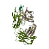
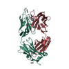
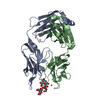
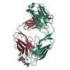
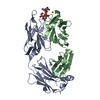
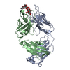
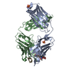
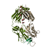
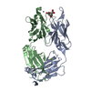
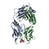
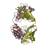
| Entry | Database: PDB / ID: 5ock | ||||||||||||
|---|---|---|---|---|---|---|---|---|---|---|---|---|---|
| Title | Crystal structure of ACPA E4 in complex with CEP1 | ||||||||||||
 Components Components |
| ||||||||||||
 Keywords Keywords | IMMUNE SYSTEM / anti-citrullinated protein antibody Fab fragment | ||||||||||||
| Function / homology |  Function and homology information Function and homology informationManipulation of host energy metabolism / positive regulation of plasminogen activation / phosphopyruvate hydratase / phosphopyruvate hydratase complex / phosphopyruvate hydratase activity / negative regulation of hypoxia-induced intrinsic apoptotic signaling pathway / positive regulation of muscle contraction / Gluconeogenesis / Glycolysis / M band ...Manipulation of host energy metabolism / positive regulation of plasminogen activation / phosphopyruvate hydratase / phosphopyruvate hydratase complex / phosphopyruvate hydratase activity / negative regulation of hypoxia-induced intrinsic apoptotic signaling pathway / positive regulation of muscle contraction / Gluconeogenesis / Glycolysis / M band / canonical glycolysis / nuclear outer membrane / positive regulation of ATP biosynthetic process / transcription corepressor binding / glycolytic process / gluconeogenesis / RNA polymerase II transcription regulatory region sequence-specific DNA binding / negative regulation of cell growth / DNA-binding transcription repressor activity, RNA polymerase II-specific / response to virus / transcription corepressor activity / GTPase binding / cell cortex / cadherin binding / negative regulation of DNA-templated transcription / magnesium ion binding / cell surface / negative regulation of transcription by RNA polymerase II / protein homodimerization activity / : / RNA binding / extracellular exosome / membrane / nucleus / plasma membrane / cytoplasm / cytosol Similarity search - Function | ||||||||||||
| Biological species |  Homo sapiens (human) Homo sapiens (human) | ||||||||||||
| Method |  X-RAY DIFFRACTION / SYNCHROTRON / MOLECULAR REPLACEMENT / Resolution: 1.6 Å X-RAY DIFFRACTION / SYNCHROTRON / MOLECULAR REPLACEMENT / Resolution: 1.6 Å | ||||||||||||
 Authors Authors | Dobritzsch, D. / Ge, C. / Holmdahl, R. | ||||||||||||
| Funding support |  Sweden, 2items Sweden, 2items
| ||||||||||||
 Citation Citation | Journal: Arthritis Rheumatol / Year: 2019 Title: Structural Basis of Cross-Reactivity of Anti-Citrullinated Protein Antibodies. Authors: Ge, C. / Xu, B. / Liang, B. / Lonnblom, E. / Lundstrom, S.L. / Zubarev, R.A. / Ayoglu, B. / Nilsson, P. / Skogh, T. / Kastbom, A. / Malmstrom, V. / Klareskog, L. / Toes, R.E.M. / Rispens, T. ...Authors: Ge, C. / Xu, B. / Liang, B. / Lonnblom, E. / Lundstrom, S.L. / Zubarev, R.A. / Ayoglu, B. / Nilsson, P. / Skogh, T. / Kastbom, A. / Malmstrom, V. / Klareskog, L. / Toes, R.E.M. / Rispens, T. / Dobritzsch, D. / Holmdahl, R. | ||||||||||||
| History |
|
- Structure visualization
| Structure viewer | Molecule: MolmilJmol/JSmol |
|---|
- Downloads & links
-Download
| PDBx/mmCIF format | 5ock.cif.gz | 114.3 KB | Display | PDBx/mmCIF format |
|---|---|---|---|---|
| PDB format | pdb5ock.ent.gz | 85.4 KB | Display | PDB format |
| PDBx/mmJSON format | 5ock.json.gz | Tree view | PDBx/mmJSON format | |
| Others |  Other downloads Other downloads |
-Validation report
| Arichive directory | https://data.pdbj.org/pub/pdb/validation_reports/oc/5ockftp://data.pdbj.org/pub/pdb/validation_reports/oc/5ock | HTTPS FTP |
|---|
-Related structure data
| Related structure data |  5ocxC  5ocyC  5od0C  5od8C  5odbC  3lmjS 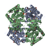 4m1qS S: Starting model for refinement C: citing same article ( |
|---|---|
| Similar structure data |

-Links
 PDBj
PDBj


- Assembly
| Deposited unit | 
| ||||||||
|---|---|---|---|---|---|---|---|---|---|
| 1 |
| ||||||||
| Unit cell |
|
-Components
| #1: Antibody | Mass: 23622.998 Da / Num. of mol.: 1 Source method: isolated from a genetically manipulated source Details: B cells of a rheumatoid arthritis patient / Source: (gene. exp.) Homo sapiens (human) / Cell line (production host): Expi293F (TM) / Production host: Homo sapiens (human) |
|---|---|
| #2: Antibody | Mass: 23437.303 Da / Num. of mol.: 1 Source method: isolated from a genetically manipulated source Details: B cells of a rheumatoid arthritis patient / Source: (gene. exp.) Homo sapiens (human) / Cell (production host): Expi293F (TM) / Cell line (production host): Expi293F (TM) / Production host: Homo sapiens (human) |
| #3: Protein/peptide | Mass: 2421.771 Da / Num. of mol.: 1 / Source method: obtained synthetically Details: Biotin is attached to the aminohexanoic acid at the N-terminus Glu7 and Ile8 not modeled due to poor definition in electron density Source: (synth.) Homo sapiens (human) / References: UniProt: P06733*PLUS |
| #4: Water | ChemComp-HOH /  Mass: 18.015 Da / Num. of mol.: 456 / Source method: isolated from a natural source / Formula: H2O Mass: 18.015 Da / Num. of mol.: 456 / Source method: isolated from a natural source / Formula: H2O |
| Has protein modification | Y |
-Experimental details
-Experiment
| Experiment | Method: X-RAY DIFFRACTION / Number of used crystals: 1 |
|---|
- Sample preparation
| Crystal | Density Matthews: 2.07 Å3/Da / Density % sol: 40.51 % |
|---|---|
| Crystal grow | Temperature: 293 K / Method: vapor diffusion, sitting drop / Details: 20% PEG3350, 0.2M ammonium citrate |
-Data collection
| Diffraction | Mean temperature: 100 K |
|---|---|
| Diffraction source | Source: SYNCHROTRON / Site: Diamond  / Beamline: I24 / Wavelength: 0.9686 Å / Beamline: I24 / Wavelength: 0.9686 Å |
| Detector | Type: DECTRIS PILATUS3 S 6M / Detector: PIXEL / Date: Feb 20, 2016 |
| Radiation | Protocol: SINGLE WAVELENGTH / Monochromatic (M) / Laue (L): M / Scattering type: x-ray |
| Radiation wavelength | Wavelength: 0.9686 Å / Relative weight: 1 |
| Reflection | Resolution: 1.6→45.63 Å / Num. obs: 51683 / % possible obs: 98.4 % / Redundancy: 3.7 % / Biso Wilson estimate: 12.5 Å2 / CC1/2: 0.961 / Rmerge(I) obs: 0.112 / Rpim(I) all: 0.089 / Net I/σ(I): 6.7 |
| Reflection shell | Resolution: 1.6→1.63 Å / Redundancy: 3.7 % / Rmerge(I) obs: 0.661 / Mean I/σ(I) obs: 1.8 / Num. unique obs: 2575 / CC1/2: 0.498 / Rpim(I) all: 0.538 / % possible all: 98.4 |
- Processing
| Software |
| ||||||||||||||||||||||||||||||||||||||||||||||||||||||||||||||||||||||||||||||||||||||||||||||||||||||||||||||||||||||||||||||||||||||||||||||||||||||||||||||||||||||||||||||||||||||
|---|---|---|---|---|---|---|---|---|---|---|---|---|---|---|---|---|---|---|---|---|---|---|---|---|---|---|---|---|---|---|---|---|---|---|---|---|---|---|---|---|---|---|---|---|---|---|---|---|---|---|---|---|---|---|---|---|---|---|---|---|---|---|---|---|---|---|---|---|---|---|---|---|---|---|---|---|---|---|---|---|---|---|---|---|---|---|---|---|---|---|---|---|---|---|---|---|---|---|---|---|---|---|---|---|---|---|---|---|---|---|---|---|---|---|---|---|---|---|---|---|---|---|---|---|---|---|---|---|---|---|---|---|---|---|---|---|---|---|---|---|---|---|---|---|---|---|---|---|---|---|---|---|---|---|---|---|---|---|---|---|---|---|---|---|---|---|---|---|---|---|---|---|---|---|---|---|---|---|---|---|---|---|---|
| Refinement | Method to determine structure: MOLECULAR REPLACEMENT Starting model: 4m1q, 3lmj Resolution: 1.6→45.63 Å / Cor.coef. Fo:Fc: 0.958 / Cor.coef. Fo:Fc free: 0.939 / SU B: 2.382 / SU ML: 0.081 / Cross valid method: THROUGHOUT / ESU R: 0.104 / ESU R Free: 0.102 / Details: HYDROGENS HAVE BEEN ADDED IN THE RIDING POSITIONS
| ||||||||||||||||||||||||||||||||||||||||||||||||||||||||||||||||||||||||||||||||||||||||||||||||||||||||||||||||||||||||||||||||||||||||||||||||||||||||||||||||||||||||||||||||||||||
| Solvent computation | Ion probe radii: 0.8 Å / Shrinkage radii: 0.8 Å / VDW probe radii: 1.2 Å | ||||||||||||||||||||||||||||||||||||||||||||||||||||||||||||||||||||||||||||||||||||||||||||||||||||||||||||||||||||||||||||||||||||||||||||||||||||||||||||||||||||||||||||||||||||||
| Displacement parameters | Biso mean: 16.425 Å2
| ||||||||||||||||||||||||||||||||||||||||||||||||||||||||||||||||||||||||||||||||||||||||||||||||||||||||||||||||||||||||||||||||||||||||||||||||||||||||||||||||||||||||||||||||||||||
| Refinement step | Cycle: 1 / Resolution: 1.6→45.63 Å
| ||||||||||||||||||||||||||||||||||||||||||||||||||||||||||||||||||||||||||||||||||||||||||||||||||||||||||||||||||||||||||||||||||||||||||||||||||||||||||||||||||||||||||||||||||||||
| Refine LS restraints |
|