 Movie
Movie Controller


+ Open data

- Basic information
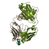
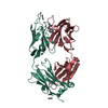
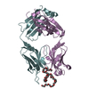
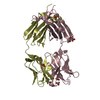
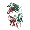
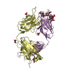
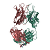
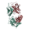
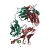
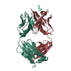
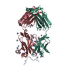
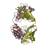
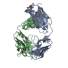
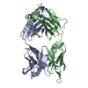
| Entry | Database: PDB / ID: 1opg | ||||||
|---|---|---|---|---|---|---|---|
| Title | OPG2 FAB FRAGMENT | ||||||
 Components Components |
| ||||||
 Keywords Keywords | IMMUNOGLOBULIN / CELL ADHESION MOLECULE | ||||||
| Function / homology |  Function and homology information Function and homology information | ||||||
| Biological species |  Homo sapiens (human) Homo sapiens (human) | ||||||
| Method |  X-RAY DIFFRACTION / Resolution: 2 Å X-RAY DIFFRACTION / Resolution: 2 Å | ||||||
 Authors Authors | Kodandapani, R. / Veerapandian, B. / Ely, K.R. | ||||||
 Citation Citation | Journal: J.Biol.Chem. / Year: 1995 Title: Crystal structure of the OPG2 Fab. An antireceptor antibody that mimics an RGD cell adhesion site. Authors: Kodandapani, R. / Veerapandian, B. / Kunicki, T.J. / Ely, K.R. #1: Journal: Acta Crystallogr.,Sect.D / Year: 1992Title: Crystallization of Opg2 Fab Fragment: A Fibrinogen Mimic with Specificity for the Platelet Glycoprotein Authors: Celikel, R. / Williamson, M.M. / Kunicki, T.J. / Ely, K.R. | ||||||
| History |
|
- Structure visualization
| Structure viewer | Molecule: MolmilJmol/JSmol |
|---|
- Downloads & links
-Download
| PDBx/mmCIF format | 1opg.cif.gz | 115 KB | Display | PDBx/mmCIF format |
|---|---|---|---|---|
| PDB format | pdb1opg.ent.gz | 86.3 KB | Display | PDB format |
| PDBx/mmJSON format | 1opg.json.gz | Tree view | PDBx/mmJSON format | |
| Others |  Other downloads Other downloads |
-Validation report
| Arichive directory | https://data.pdbj.org/pub/pdb/validation_reports/op/1opgftp://data.pdbj.org/pub/pdb/validation_reports/op/1opg | HTTPS FTP |
|---|
-Related structure data
| Similar structure data |
|---|


-Links
 PDBj
PDBj

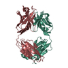
- Assembly
| Deposited unit | 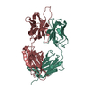
| ||||||||
|---|---|---|---|---|---|---|---|---|---|
| 1 |
| ||||||||
| Unit cell |
| ||||||||
| Atom site foot note | 1: CIS PROLINE - PRO L 8 / 2: CIS PROLINE - PRO L 95 / 3: CIS PROLINE - PRO L 141 / 4: CIS PROLINE - PRO H 159 / 5: CIS PROLINE - PRO H 161 |
-Components
| #1: Antibody | Mass: 23549.811 Da / Num. of mol.: 1 / Source method: isolated from a natural source / Source: (natural) Homo sapiens (human) / Tissue: PLATELET |
|---|---|
| #2: Antibody | Mass: 24508.473 Da / Num. of mol.: 1 / Source method: isolated from a natural source / Source: (natural) Homo sapiens (human) / Tissue: PLATELET / References: UniProt: P01868 |
| #3: Water | ChemComp-HOH /  Mass: 18.015 Da / Num. of mol.: 488 / Source method: isolated from a natural source / Formula: H2O Mass: 18.015 Da / Num. of mol.: 488 / Source method: isolated from a natural source / Formula: H2O |
| Has protein modification | Y |
-Experimental details
-Experiment
| Experiment | Method: X-RAY DIFFRACTION |
|---|
- Sample preparation
| Crystal | Density Matthews: 2.18 Å3/Da / Density % sol: 43.53 % | ||||||||||||||||||||||||
|---|---|---|---|---|---|---|---|---|---|---|---|---|---|---|---|---|---|---|---|---|---|---|---|---|---|
| Crystal grow | *PLUS Temperature: 22 ℃ / Method: vapor diffusion, hanging dropDetails: Celikel, R., (1993) Acta Crystallogr.,Sect.D, 49, 421. | ||||||||||||||||||||||||
| Components of the solutions | *PLUS
|
-Data collection
| Detector | Date: Mar 12, 1992 |
|---|---|
| Radiation | Monochromatic (M) / Laue (L): M / Scattering type: x-ray |
| Radiation wavelength | Relative weight: 1 |
| Reflection | Num. obs: 28703 / % possible obs: 99 % / Redundancy: 3.2 % / Rmerge(I) obs: 0.044 |
| Reflection | *PLUS Highest resolution: 2 Å / Num. measured all: 89211 / Rmerge(I) obs: 0.044 |
- Processing
| Software |
| ||||||||||||||||||||||||||||||||||||||||||||||||||||||||||||||||||||||||||||||||||||
|---|---|---|---|---|---|---|---|---|---|---|---|---|---|---|---|---|---|---|---|---|---|---|---|---|---|---|---|---|---|---|---|---|---|---|---|---|---|---|---|---|---|---|---|---|---|---|---|---|---|---|---|---|---|---|---|---|---|---|---|---|---|---|---|---|---|---|---|---|---|---|---|---|---|---|---|---|---|---|---|---|---|---|---|---|---|
| Refinement | Resolution: 2→10 Å / σ(F): 3 /
| ||||||||||||||||||||||||||||||||||||||||||||||||||||||||||||||||||||||||||||||||||||
| Displacement parameters | Biso mean: 30.5 Å2 | ||||||||||||||||||||||||||||||||||||||||||||||||||||||||||||||||||||||||||||||||||||
| Refinement step | Cycle: LAST / Resolution: 2→10 Å
| ||||||||||||||||||||||||||||||||||||||||||||||||||||||||||||||||||||||||||||||||||||
| Refine LS restraints |
| ||||||||||||||||||||||||||||||||||||||||||||||||||||||||||||||||||||||||||||||||||||
| Software | *PLUS Name: PROLSQ / Classification: refinement | ||||||||||||||||||||||||||||||||||||||||||||||||||||||||||||||||||||||||||||||||||||
| Refinement | *PLUS Rfactor obs: 0.16 | ||||||||||||||||||||||||||||||||||||||||||||||||||||||||||||||||||||||||||||||||||||
| Solvent computation | *PLUS | ||||||||||||||||||||||||||||||||||||||||||||||||||||||||||||||||||||||||||||||||||||
| Displacement parameters | *PLUS |
