 Movie
Movie Controller


+ Open data

- Basic information
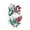
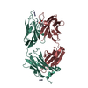
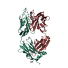
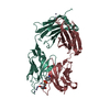
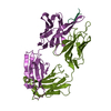
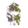
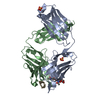
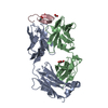
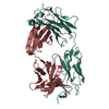
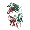
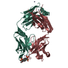
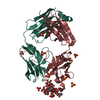
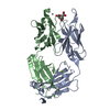
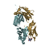
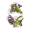
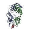
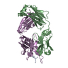
| Entry | Database: PDB / ID: 1kn4 | ||||||
|---|---|---|---|---|---|---|---|
| Title | CATALYTIC ANTIBODY D2.3 COMPLEX | ||||||
 Components Components |
| ||||||
 Keywords Keywords | IMMUNE SYSTEM / ABZYME / TRANSITION STATE ANALOG | ||||||
| Function / homology |  Function and homology information Function and homology informationFc-gamma receptor I complex binding / immunoglobulin complex, circulating / immunoglobulin receptor binding / IgG immunoglobulin complex / complement activation, classical pathway / immunoglobulin complex / antigen binding / antibacterial humoral response / adaptive immune response / : ...Fc-gamma receptor I complex binding / immunoglobulin complex, circulating / immunoglobulin receptor binding / IgG immunoglobulin complex / complement activation, classical pathway / immunoglobulin complex / antigen binding / antibacterial humoral response / adaptive immune response / : / extracellular region / metal ion binding / plasma membrane Similarity search - Function | ||||||
| Biological species |  | ||||||
| Method |  X-RAY DIFFRACTION / SYNCHROTRON / MOLECULAR REPLACEMENT / Resolution: 1.9 Å X-RAY DIFFRACTION / SYNCHROTRON / MOLECULAR REPLACEMENT / Resolution: 1.9 Å | ||||||
 Authors Authors | Gigant, B. / Knossow, M. | ||||||
 Citation Citation | Journal: J.Am.Chem.Soc. / Year: 2002 Title: Remarkable remote chiral recognition in a reaction mediated by a catalytic antibody. Authors: D'Souza, L.J. / Gigant, B. / Knossow, M. / Green, B.S. | ||||||
| History |
| ||||||
| Remark 600 | HETEROGEN PDE IS PARA-NITROPHENYL PHOSPHONOBUTANOYL D-ALANINE (NO2-C6H5-O-PO2-CH2-CH2-CH2-CO-NH-CH(CH3)-COOH) | ||||||
| Remark 999 | SEQUENCE THE SEQUENCES OF THE CONSTANT DOMAINS OF THE HEAVY CHAINS. (RESIDUES H106 - H223) AND OF ...SEQUENCE THE SEQUENCES OF THE CONSTANT DOMAINS OF THE HEAVY CHAINS. (RESIDUES H106 - H223) AND OF THE LIGHT CHAINS (RESIDUES L107 - L214) HAVE NOT BEEN DETERMINED FOR THIS IMMUNOGLOBULIN. THEY HAVE BEEN ASSIGNED THE CONSENSUS SEQUENCE FOR THE CONSTANT DOMAIN OF MOUSE KAPPA LIGHT CHAIN AND FOR THE FIRST CONSTANT DOMAIN OF MOUSE GROUP 2A HEAVY CHAINS. |
- Structure visualization
| Structure viewer | Molecule: MolmilJmol/JSmol |
|---|
- Downloads & links
-Download
| PDBx/mmCIF format | 1kn4.cif.gz | 103.8 KB | Display | PDBx/mmCIF format |
|---|---|---|---|---|
| PDB format | pdb1kn4.ent.gz | 78 KB | Display | PDB format |
| PDBx/mmJSON format | 1kn4.json.gz | Tree view | PDBx/mmJSON format | |
| Others |  Other downloads Other downloads |
-Validation report
| Arichive directory | https://data.pdbj.org/pub/pdb/validation_reports/kn/1kn4ftp://data.pdbj.org/pub/pdb/validation_reports/kn/1kn4 | HTTPS FTP |
|---|
-Related structure data
| Related structure data | 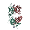 1kn2C 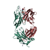 1yecS S: Starting model for refinement C: citing same article ( |
|---|---|
| Similar structure data |


-Links
 PDBj
PDBj


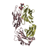
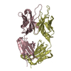
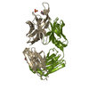
- Assembly
| Deposited unit | 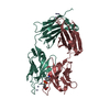
| ||||||||
|---|---|---|---|---|---|---|---|---|---|
| 1 |
| ||||||||
| 2 | 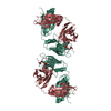
| ||||||||
| 3 | 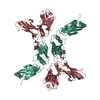
| ||||||||
| 4 | 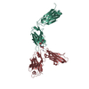
| ||||||||
| Unit cell |
|
-Components
| #1: Antibody | Mass: 24019.842 Da / Num. of mol.: 1 / Source method: isolated from a natural source / Source: (natural) | ||||||
|---|---|---|---|---|---|---|---|
| #2: Antibody | Mass: 24189.217 Da / Num. of mol.: 1 / Source method: isolated from a natural source / Source: (natural) | ||||||
| #3: Chemical | ChemComp-ZN /   Mass: 65.409 Da / Num. of mol.: 7 / Source method: obtained synthetically / Formula: Zn Mass: 65.409 Da / Num. of mol.: 7 / Source method: obtained synthetically / Formula: Zn#4: Chemical | ChemComp-PDE / | 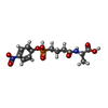  Mass: 360.256 Da / Num. of mol.: 1 / Source method: obtained synthetically / Formula: C13H17N2O8P Mass: 360.256 Da / Num. of mol.: 1 / Source method: obtained synthetically / Formula: C13H17N2O8P#5: Water | ChemComp-HOH / |  Mass: 18.015 Da / Num. of mol.: 151 / Source method: isolated from a natural source / Formula: H2O Mass: 18.015 Da / Num. of mol.: 151 / Source method: isolated from a natural source / Formula: H2OHas protein modification | Y | |
-Experimental details
-Experiment
| Experiment | Method: X-RAY DIFFRACTION / Number of used crystals: 1 |
|---|
- Sample preparation
| Crystal | Density Matthews: 2.91 Å3/Da / Density % sol: 57 % | |||||||||||||||||||||||||
|---|---|---|---|---|---|---|---|---|---|---|---|---|---|---|---|---|---|---|---|---|---|---|---|---|---|---|
| Crystal grow | Method: vapor diffusion, hanging drop / pH: 7 Details: PEG600, ZnSO4, Cacodylate, pH 7.00, VAPOR DIFFUSION, HANGING DROP | |||||||||||||||||||||||||
| Crystal grow | *PLUS Temperature: 18 ℃ / pH: 7.5 / Details: Gigant, B., (1998) J. Mol. Biol., 284, 741. | |||||||||||||||||||||||||
| Components of the solutions | *PLUS
|
-Data collection
| Diffraction | Mean temperature: 278 K |
|---|---|
| Diffraction source | Source: SYNCHROTRON / Site: LURE  / Beamline: DW32 / Wavelength: 0.963 / Beamline: DW32 / Wavelength: 0.963 |
| Detector | Type: MARRESEARCH / Detector: IMAGE PLATE / Date: Dec 8, 1998 / Details: BENT MIRROR |
| Radiation | Monochromator: GRAPHITE / Protocol: SINGLE WAVELENGTH / Monochromatic (M) / Laue (L): M / Scattering type: x-ray |
| Radiation wavelength | Wavelength: 0.963 Å / Relative weight: 1 |
| Reflection | Resolution: 1.9→20 Å / Num. all: 43212 / Num. obs: 43212 / % possible obs: 95.5 % / Observed criterion σ(F): 0 / Observed criterion σ(I): -3 / Redundancy: 2.6 % / Biso Wilson estimate: 23.8 Å2 / Rsym value: 0.07 / Net I/σ(I): 12 |
| Reflection shell | Resolution: 1.9→1.93 Å / Mean I/σ(I) obs: 2.3 / Rsym value: 0.0439 / % possible all: 97.6 |
| Reflection | *PLUS Highest resolution: 1.9 Å / Lowest resolution: 20 Å / Num. obs: 42817 / % possible obs: 94.7 % / Num. measured all: 106350 / Rmerge(I) obs: 0.085 |
| Reflection shell | *PLUS % possible obs: 98.2 % / Rmerge(I) obs: 0.342 / Mean I/σ(I) obs: 3.1 |
- Processing
| Software |
| ||||||||||||||||||||||||||||||||||||||||||||||||||||||||||||||||||||||||||||||||
|---|---|---|---|---|---|---|---|---|---|---|---|---|---|---|---|---|---|---|---|---|---|---|---|---|---|---|---|---|---|---|---|---|---|---|---|---|---|---|---|---|---|---|---|---|---|---|---|---|---|---|---|---|---|---|---|---|---|---|---|---|---|---|---|---|---|---|---|---|---|---|---|---|---|---|---|---|---|---|---|---|---|
| Refinement | Method to determine structure: MOLECULAR REPLACEMENT Starting model: 1YEC Resolution: 1.9→20 Å / Rfactor Rfree error: 0.005 / Data cutoff high absF: 1811954.89 / Data cutoff high rms absF: 1811954.89 / Data cutoff low absF: 0 / Isotropic thermal model: RESTRAINED / Cross valid method: THROUGHOUT / σ(F): 0
| ||||||||||||||||||||||||||||||||||||||||||||||||||||||||||||||||||||||||||||||||
| Displacement parameters | Biso mean: 31.8 Å2
| ||||||||||||||||||||||||||||||||||||||||||||||||||||||||||||||||||||||||||||||||
| Refine analyze |
| ||||||||||||||||||||||||||||||||||||||||||||||||||||||||||||||||||||||||||||||||
| Refinement step | Cycle: LAST / Resolution: 1.9→20 Å
| ||||||||||||||||||||||||||||||||||||||||||||||||||||||||||||||||||||||||||||||||
| Refine LS restraints |
| ||||||||||||||||||||||||||||||||||||||||||||||||||||||||||||||||||||||||||||||||
| LS refinement shell | Resolution: 1.9→1.99 Å / Rfactor Rfree error: 0.017 / Total num. of bins used: 8
| ||||||||||||||||||||||||||||||||||||||||||||||||||||||||||||||||||||||||||||||||
| Refinement | *PLUS Highest resolution: 1.9 Å / Lowest resolution: 20 Å / σ(F): 0 / % reflection Rfree: 5.1 % / Rfactor obs: 0.213 | ||||||||||||||||||||||||||||||||||||||||||||||||||||||||||||||||||||||||||||||||
| Solvent computation | *PLUS | ||||||||||||||||||||||||||||||||||||||||||||||||||||||||||||||||||||||||||||||||
| Displacement parameters | *PLUS Biso mean: 31.8 Å2 | ||||||||||||||||||||||||||||||||||||||||||||||||||||||||||||||||||||||||||||||||
| Refine LS restraints | *PLUS
| ||||||||||||||||||||||||||||||||||||||||||||||||||||||||||||||||||||||||||||||||
| LS refinement shell | *PLUS % reflection Rfree: 5.2 % / Rfactor Rwork: 0.275 / Rfactor obs: 0.275 |
