 Movie
Movie Controller

[English] 日本語
 Yorodumi
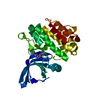
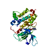
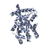
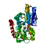
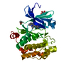
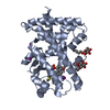
- PDB-4pf6: CRYSTAL STRUCTURE OF A TRAP PERIPLASMIC SOLUTE BINDING PROTEIN FR... -
+ Open data

- Basic information
| Entry | Database: PDB / ID: 4pf6 | ||||||
|---|---|---|---|---|---|---|---|
| Title | CRYSTAL STRUCTURE OF A TRAP PERIPLASMIC SOLUTE BINDING PROTEIN FROM ROSEOBACTER DENITRIFICANS (RD1_0742, TARGET EFI-510239) WITH BOUND 3-DEOXY-D-MANNO-OCT-2-ULOSONIC ACID (KDO) | ||||||
 Components Components | C4-dicarboxylate-binding protein | ||||||
 Keywords Keywords | TRANSPORT PROTEIN / TRAP PERIPLASMIC SOLUTE BINDING FAMILY / ENZYME FUNCTION INITIATIVE / EFI / Structural Genomics | ||||||
| Function / homology |  Function and homology information Function and homology information | ||||||
| Biological species |  Roseobacter denitrificans (bacteria) Roseobacter denitrificans (bacteria) | ||||||
| Method |  X-RAY DIFFRACTION / SYNCHROTRON / SAD / Resolution: 1.75 Å X-RAY DIFFRACTION / SYNCHROTRON / SAD / Resolution: 1.75 Å | ||||||
 Authors Authors | Vetting, M.W. / Al Obaidi, N.F. / Morisco, L.L. / Wasserman, S.R. / Stead, M. / Attonito, J.D. / Scott Glenn, A. / Chowdhury, S. / Evans, B. / Hillerich, B. ...Vetting, M.W. / Al Obaidi, N.F. / Morisco, L.L. / Wasserman, S.R. / Stead, M. / Attonito, J.D. / Scott Glenn, A. / Chowdhury, S. / Evans, B. / Hillerich, B. / Love, J. / Seidel, R.D. / Whalen, K.L. / Gerlt, J.A. / Almo, S.C. / Enzyme Function Initiative (EFI) | ||||||
| Funding support |  United States, 1items United States, 1items
| ||||||
 Citation Citation | Journal: Biochemistry / Year: 2015 Title: Experimental strategies for functional annotation and metabolism discovery: targeted screening of solute binding proteins and unbiased panning of metabolomes. Authors: Vetting, M.W. / Al-Obaidi, N. / Zhao, S. / San Francisco, B. / Kim, J. / Wichelecki, D.J. / Bouvier, J.T. / Solbiati, J.O. / Vu, H. / Zhang, X. / Rodionov, D.A. / Love, J.D. / Hillerich, B.S. ...Authors: Vetting, M.W. / Al-Obaidi, N. / Zhao, S. / San Francisco, B. / Kim, J. / Wichelecki, D.J. / Bouvier, J.T. / Solbiati, J.O. / Vu, H. / Zhang, X. / Rodionov, D.A. / Love, J.D. / Hillerich, B.S. / Seidel, R.D. / Quinn, R.J. / Osterman, A.L. / Cronan, J.E. / Jacobson, M.P. / Gerlt, J.A. / Almo, S.C. | ||||||
| History |
|
- Structure visualization
| Structure viewer | Molecule: MolmilJmol/JSmol |
|---|
- Downloads & links
-Download
| PDBx/mmCIF format | 4pf6.cif.gz | 195.9 KB | Display | PDBx/mmCIF format |
|---|---|---|---|---|
| PDB format | pdb4pf6.ent.gz | 158.8 KB | Display | PDB format |
| PDBx/mmJSON format | 4pf6.json.gz | Tree view | PDBx/mmJSON format | |
| Others |  Other downloads Other downloads |
-Validation report
| Arichive directory | https://data.pdbj.org/pub/pdb/validation_reports/pf/4pf6ftp://data.pdbj.org/pub/pdb/validation_reports/pf/4pf6 | HTTPS FTP |
|---|
-Related structure data
| Related structure data | 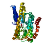 4ln5C  4mcoC 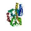 4mevC  4mhfC 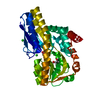 4mijC 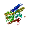 4mncC 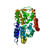 4mniC 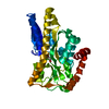 4mx6C 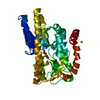 4n15C 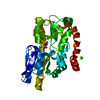 4n17C  4n4uC 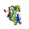 4n6dC 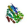 4n6kC 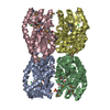 4n8gC  4n8yC 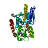 4n91C 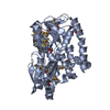 4napC 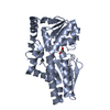 4nf0C 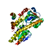 4ng7C 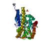 4nguC 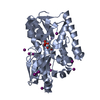 4nhbC 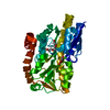 4nn3C 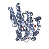 4nq8C 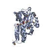 4nx1C 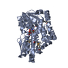 4o7mC 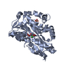 4o8mC  4o94C  4oa4C  4oanC 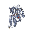 4ovpC  4ovqC  4ovrC  4ovsC 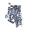 4ovtC 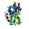 4p1eC 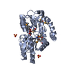 4p1lC 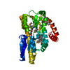 4p3lC 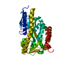 4p47C 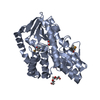 4p56C 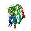 4p8bC 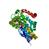 4p9kC  4pafC  4paiC 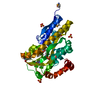 4pakC 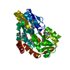 4pbhC 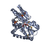 4pbqC  4pc9C  4pcdC  4pddC  4pdhC 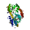 4pe3C 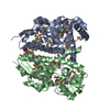 4petC 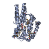 4pf8C 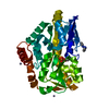 4pfbC 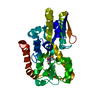 4pfiC 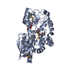 4pfrC 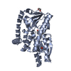 4pgnC 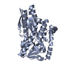 4pgpC 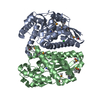 4uabC C: citing same article ( |
|---|---|
| Similar structure data | |
| Other databases |

-Links
 PDBj
PDBj- Assembly
| Deposited unit | 
| ||||||||
|---|---|---|---|---|---|---|---|---|---|
| 1 |
| ||||||||
| Unit cell |
| ||||||||
| Components on special symmetry positions |
| ||||||||
| Details | biological unit is a monomer |
-Components
| #1: Protein | Mass: 36671.328 Da / Num. of mol.: 1 / Fragment: UNP residues 25-331 Source method: isolated from a genetically manipulated source Source: (gene. exp.) Roseobacter denitrificans (bacteria) / Strain: ATCC 33942 / OCh 114 / Gene: RD1_0742 / Plasmid: pET / Production host: | ||||
|---|---|---|---|---|---|
| #2: Sugar | ChemComp-KDO / 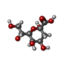  Type: D-saccharide, alpha linking / Mass: 238.192 Da / Num. of mol.: 1 Type: D-saccharide, alpha linking / Mass: 238.192 Da / Num. of mol.: 1Source method: isolated from a genetically manipulated source Formula: C8H14O8 | ||||
| #3: Chemical | ChemComp-PGE /   Mass: 150.173 Da / Num. of mol.: 1 / Source method: obtained synthetically / Formula: C6H14O4 Mass: 150.173 Da / Num. of mol.: 1 / Source method: obtained synthetically / Formula: C6H14O4 | ||||
| #4: Chemical |   Mass: 96.063 Da / Num. of mol.: 2 / Source method: obtained synthetically / Formula: SO4 Mass: 96.063 Da / Num. of mol.: 2 / Source method: obtained synthetically / Formula: SO4#5: Water | ChemComp-HOH / |  Mass: 18.015 Da / Num. of mol.: 434 / Source method: isolated from a natural source / Formula: H2O Mass: 18.015 Da / Num. of mol.: 434 / Source method: isolated from a natural source / Formula: H2OHas protein modification | Y | |
-Experimental details
-Experiment
| Experiment | Method: X-RAY DIFFRACTION / Number of used crystals: 1 |
|---|
- Sample preparation
| Crystal | Density Matthews: 3.17 Å3/Da / Density % sol: 61.15 % |
|---|---|
| Crystal grow | Temperature: 298 K / Method: vapor diffusion, sitting drop / pH: 6.5 Details: Protein (31.6 mg/ml, 10 mM HEPES pH 7.5, 5 mM DTT); Reservoir (0.2 M Sodium Chloride, 0.1 M Sodium Cacodylate pH 6.5, 2 M Ammonium Sulfate ); Cryoprotection (80% LiSO4 (2 M) + 20% reservoir |
-Data collection
| Diffraction | Mean temperature: 100 K | ||||||||||||||||||||||||
|---|---|---|---|---|---|---|---|---|---|---|---|---|---|---|---|---|---|---|---|---|---|---|---|---|---|
| Diffraction source | Source: SYNCHROTRON / Site: APS / Beamline: 31-ID / Wavelength: 0.9793 Å | ||||||||||||||||||||||||
| Detector | Type: RAYONIX MX225HE / Detector: CCD / Date: Apr 9, 2014 / Details: MIRRORS | ||||||||||||||||||||||||
| Radiation | Monochromator: GRAPHITE / Protocol: SINGLE WAVELENGTH / Monochromatic (M) / Laue (L): M / Scattering type: x-ray | ||||||||||||||||||||||||
| Radiation wavelength | Wavelength: 0.9793 Å / Relative weight: 1 | ||||||||||||||||||||||||
| Reflection | Resolution: 1.7→159.21 Å / Num. obs: 52900 / % possible obs: 100 % / Redundancy: 20.3 % / Biso Wilson estimate: 19.52 Å2 / Rmerge(I) obs: 0.104 / Rpim(I) all: 0.023 / Net I/σ(I): 25 / Num. measured all: 1071804 / Scaling rejects: 27 | ||||||||||||||||||||||||
| Reflection shell | Diffraction-ID: 1 / Rejects: _
|
- Processing
| Software |
| ||||||||||||||||||||||||||||||||||||||||||||||||||||||||||||||||||||||||||||||||||||||||||||||||||||||||||||||||||||||||||||||||||||||||||||||||||||||||||||||||||||||||||||||||||||||||||||||||||||||||||||||||||||||||||||||||||||||||||||||||||||||||||
|---|---|---|---|---|---|---|---|---|---|---|---|---|---|---|---|---|---|---|---|---|---|---|---|---|---|---|---|---|---|---|---|---|---|---|---|---|---|---|---|---|---|---|---|---|---|---|---|---|---|---|---|---|---|---|---|---|---|---|---|---|---|---|---|---|---|---|---|---|---|---|---|---|---|---|---|---|---|---|---|---|---|---|---|---|---|---|---|---|---|---|---|---|---|---|---|---|---|---|---|---|---|---|---|---|---|---|---|---|---|---|---|---|---|---|---|---|---|---|---|---|---|---|---|---|---|---|---|---|---|---|---|---|---|---|---|---|---|---|---|---|---|---|---|---|---|---|---|---|---|---|---|---|---|---|---|---|---|---|---|---|---|---|---|---|---|---|---|---|---|---|---|---|---|---|---|---|---|---|---|---|---|---|---|---|---|---|---|---|---|---|---|---|---|---|---|---|---|---|---|---|---|---|---|---|---|---|---|---|---|---|---|---|---|---|---|---|---|---|---|---|---|---|---|---|---|---|---|---|---|---|---|---|---|---|---|---|---|---|---|---|---|---|---|---|---|---|---|---|---|---|---|
| Refinement | Method to determine structure: SAD / Resolution: 1.75→47.93 Å / SU ML: 0.12 / Cross valid method: FREE R-VALUE / σ(F): 1.33 / Phase error: 14.46 / Stereochemistry target values: ML
| ||||||||||||||||||||||||||||||||||||||||||||||||||||||||||||||||||||||||||||||||||||||||||||||||||||||||||||||||||||||||||||||||||||||||||||||||||||||||||||||||||||||||||||||||||||||||||||||||||||||||||||||||||||||||||||||||||||||||||||||||||||||||||
| Solvent computation | Shrinkage radii: 0.9 Å / VDW probe radii: 1.11 Å / Solvent model: FLAT BULK SOLVENT MODEL | ||||||||||||||||||||||||||||||||||||||||||||||||||||||||||||||||||||||||||||||||||||||||||||||||||||||||||||||||||||||||||||||||||||||||||||||||||||||||||||||||||||||||||||||||||||||||||||||||||||||||||||||||||||||||||||||||||||||||||||||||||||||||||
| Displacement parameters | Biso mean: 23.04 Å2 | ||||||||||||||||||||||||||||||||||||||||||||||||||||||||||||||||||||||||||||||||||||||||||||||||||||||||||||||||||||||||||||||||||||||||||||||||||||||||||||||||||||||||||||||||||||||||||||||||||||||||||||||||||||||||||||||||||||||||||||||||||||||||||
| Refinement step | Cycle: final / Resolution: 1.75→47.93 Å
| ||||||||||||||||||||||||||||||||||||||||||||||||||||||||||||||||||||||||||||||||||||||||||||||||||||||||||||||||||||||||||||||||||||||||||||||||||||||||||||||||||||||||||||||||||||||||||||||||||||||||||||||||||||||||||||||||||||||||||||||||||||||||||
| Refine LS restraints |
| ||||||||||||||||||||||||||||||||||||||||||||||||||||||||||||||||||||||||||||||||||||||||||||||||||||||||||||||||||||||||||||||||||||||||||||||||||||||||||||||||||||||||||||||||||||||||||||||||||||||||||||||||||||||||||||||||||||||||||||||||||||||||||
| LS refinement shell |
| ||||||||||||||||||||||||||||||||||||||||||||||||||||||||||||||||||||||||||||||||||||||||||||||||||||||||||||||||||||||||||||||||||||||||||||||||||||||||||||||||||||||||||||||||||||||||||||||||||||||||||||||||||||||||||||||||||||||||||||||||||||||||||
| Refinement TLS params. | Method: refined / Refine-ID: X-RAY DIFFRACTION
| ||||||||||||||||||||||||||||||||||||||||||||||||||||||||||||||||||||||||||||||||||||||||||||||||||||||||||||||||||||||||||||||||||||||||||||||||||||||||||||||||||||||||||||||||||||||||||||||||||||||||||||||||||||||||||||||||||||||||||||||||||||||||||
| Refinement TLS group |
|
