- Database of articles cited by EMDB/PDB/SASBDB data -
+
Search query
-
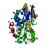
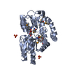
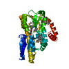
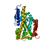
Structure paper
Title
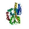
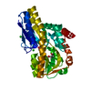
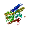
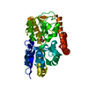
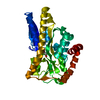
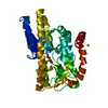
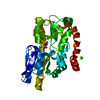
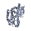
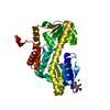
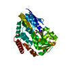
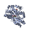
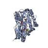
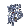
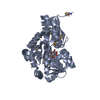
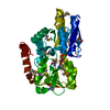
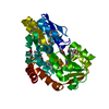
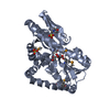
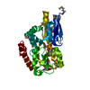
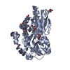
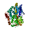
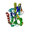
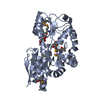
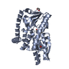
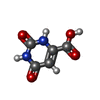
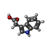
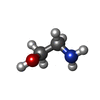
Experimental strategies for functional annotation and metabolism discovery: targeted screening of solute binding proteins and unbiased panning of metabolomes.
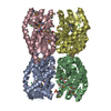
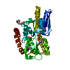
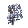
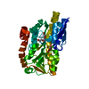
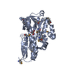
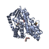
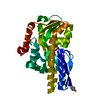
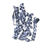
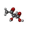
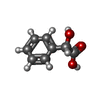
PDB-4ln5: Crystal structure of a trap periplasmic solute binding protein from burkholderia ambifaria (Bamb_6123), TARGET EFI-510059, with bound glycerol and chloride ion Method: X-RAY DIFFRACTION / Resolution: 2.1 Å
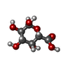
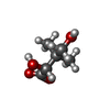
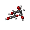
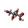
PDB-4mco: Crystal structure of a TRAP periplasmic solute binding protein from Rhodoferax ferrireducens (Rfer_1840), target EFI-510211, with bound malonate Method: X-RAY DIFFRACTION / Resolution: 1.6 Å
PDB-4mev: Crystal structure of a TRAP periplasmic solute binding protein from Rhodoferax ferrireducens (Rfer_1840), Target EFI-510211, with bound malonate, space group I422 Method: X-RAY DIFFRACTION / Resolution: 1.8 Å
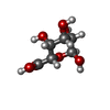
PDB-4mhf: Crystal structure of a TRAP periplasmic solute binding protein from Polaromonas sp. JS666 (Bpro_3107), target EFI-510173, with bound alpha/beta D-Glucuronate, space group P21 Method: X-RAY DIFFRACTION / Resolution: 1.46 Å
PDB-4mij: Crystal structure of a Trap periplasmic solute binding protein from Polaromonas sp. JS666 (Bpro_3107), target EFI-510173, with bound alpha/beta D-Galacturonate, space group P21 Method: X-RAY DIFFRACTION / Resolution: 1.1 Å
PDB-4mnc: Crystal structure of a TRAP periplasmic solute binding protein from Polaromonas sp. JS666 (Bpro_4736), Target EFI-510156, with bound benzoyl formate, space group P21 Method: X-RAY DIFFRACTION / Resolution: 1.05 Å
PDB-4mni: Crystal structure of a TRAP periplasmic solute binding protein from Polaromonas sp. JS666 (Bpro_4736), Target EFI-510156, with bound benzoyl formate, space group P6522 Method: X-RAY DIFFRACTION / Resolution: 1.9 Å
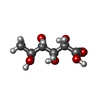
PDB-4mx6: Crystal structure of a trap periplasmic solute binding protein from shewanella oneidensis (SO_3134), target EFI-510275, with bound succinate Method: X-RAY DIFFRACTION / Resolution: 1.1 Å
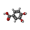
PDB-4n15: Crystal structure of a TRAP periplasmic solute binding protein from Burkholderia ambifaria (BAM_6123), Target EFI-510059, with bound beta-D-glucuronate Method: X-RAY DIFFRACTION / Resolution: 1.651 Å
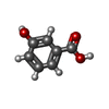
PDB-4n17: Crystal structure of a TRAP periplasmic solute binding protein from Burkholderia ambifaria (BAM_6123), Target EFI-510059, With bound beta-D-galacturonate Method: X-RAY DIFFRACTION / Resolution: 1.501 Å
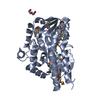
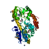
PDB-4n4u: Crystal structure of ABC transporter solute binding protein BB0719 from Bordetella bronchiseptica RB50, TARGET EFI-510049 Method: X-RAY DIFFRACTION / Resolution: 1.57 Å
PDB-4n6d: Crystal structure of a TRAP periplasmic solute binding protein from Desulfovibrio salexigens DSM2638 (Desal_3247), Target EFI-510112, phased with I3C, open complex, C-terminus of symmetry mate bound in ligand binding site Method: X-RAY DIFFRACTION / Resolution: 1.701 Å
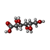
PDB-4n6k: Crystal structure of a TRAP periplasmic solute binding protein from Desulfovibrio salexigens DSM2638, Target EFI-510113 (Desal_0342), complex with diglycerolphosphate Method: X-RAY DIFFRACTION / Resolution: 1.2 Å
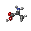
PDB-4n8g: Crystal structure of a TRAP periplasmic solute binding protein from Chromohalobacter salexigens DSM 3043 (Csal_0660), Target EFI-501075, with bound D-alanine-D-alanine Method: X-RAY DIFFRACTION / Resolution: 1.502 Å
PDB-4n8y: Crystal structure of a trap periplasmic solute binding protein from bradyrhizobium sp. btai1 b (bbta_0128), target EFI-510056 (bbta_0128), complex with alpha/beta-d-galacturonate Method: X-RAY DIFFRACTION / Resolution: 1.5 Å
PDB-4n91: Crystal structure of a trap periplasmic solute binding protein from anaerococcus prevotii dsm 20548 (Apre_1383), target EFI-510023, with bound alpha/beta d-glucuronate Method: X-RAY DIFFRACTION / Resolution: 1.7 Å
PDB-4nap: Crystal structure of a trap periplasmic solute binding protein from Desulfovibrio alaskensis G20 (DDE_0634), target EFI-510102, with bound d-tryptophan Method: X-RAY DIFFRACTION / Resolution: 2.3 Å
PDB-4nf0: CRYSTAL STRUCTURE OF A TRAP PERIPLASMIC SOLUTE BINDING PROTEIN FROM PSEUDOMONAS AERUGINOSA PAO1 (PA4616), TARGET EFI-510182, WITH BOUND L-Malate Method: X-RAY DIFFRACTION / Resolution: 1.85 Å
PDB-4ng7: Crystal structure of a TRAP periplasmic solute binding protein from Citrobacter koseri (CKO_04899), Target EFI-510094, apo, open structure Method: X-RAY DIFFRACTION / Resolution: 2.3 Å
PDB-4ngu: Crystal structure of a TRAP periplasmic solute binding protein from Desulfovibrio alaskensis G20 (Dde_1548), Target EFI-510103, with bound D-Ala-D-Ala Method: X-RAY DIFFRACTION / Resolution: 2.5 Å
PDB-4nhb: Crystal structure of a TRAP periplasmic solute binding protein from Desulfovibrio desulfuricans (Ddes_1525), Target EFI-510107, with bound sn-glycerol-3-phosphate Method: X-RAY DIFFRACTION / Resolution: 1.902 Å
PDB-4nn3: Crystal structure of a TRAP periplasmic solute binding protein from Desulfovibrio salexigens (Desal_2161), Target EFI-510109, with bound orotic acid Method: X-RAY DIFFRACTION / Resolution: 1.4 Å
PDB-4nq8: Crystal structure of a trap periplasmic solute binding protein from Bordetella bronchispeptica (bb3421), target EFI-510039, with density modeled as pantoate Method: X-RAY DIFFRACTION / Resolution: 1.5 Å
PDB-4nx1: Crystal structure of a trap periplasmic solute binding protein from Sulfitobacter sp. nas-14.1, target EFI-510292, with bound alpha-D-taluronate Method: X-RAY DIFFRACTION / Resolution: 1.6 Å
PDB-4o7m: Crystal structure of a trap periplasmic solute binding protein from shewanella loihica PV-4, target EFI-510273, with bound L-malate Method: X-RAY DIFFRACTION / Resolution: 1.5 Å
PDB-4o8m: Crystal structure of a trap periplasmic solute binding protein actinobacillus succinogenes 130z, target EFI-510004, with bound L-galactonate Method: X-RAY DIFFRACTION / Resolution: 1.7 Å
PDB-4o94: Crystal structure of a trap periplasmic solute binding protein from Rhodopseudomonas palustris HaA2 (RPB_3329), Target EFI-510223, with bound succinate Method: X-RAY DIFFRACTION / Resolution: 2 Å
PDB-4oa4: CRYSTAL STRUCTURE OF A TRAP PERIPLASMIC SOLUTE BINDING PROTEIN FROM SHEWANELLA LOIHICA PV-4 (Shew_1446), TARGET EFI-510273, WITH BOUND SUCCINATE Method: X-RAY DIFFRACTION / Resolution: 1.6 Å
PDB-4oan: Crystal structure of a TRAP periplasmic solute binding protein from rhodopseudomonas palustris HaA2 (RPB_2686), TARGET EFI-510221, with density modeled as (S)-2-hydroxy-2-methyl-3-oxobutanoate ((S)-2-Acetolactate) Method: X-RAY DIFFRACTION / Resolution: 1.35 Å
PDB-4ovp: CRYSTAL STRUCTURE OF A TRAP PERIPLASMIC SOLUTE BINDING PROTEIN FROM SULFITOBACTER sp. NAS-14.1, TARGET EFI-510292, WITH BOUND ALPHA-D-MANURONATE Method: X-RAY DIFFRACTION / Resolution: 1.7 Å
PDB-4ovq: CRYSTAL STRUCTURE OF A TRAP PERIPLASMIC SOLUTE BINDING PROTEIN FROM ROSEOBACTER DENITRIFICANS, TARGET EFI-510230, WITH BOUND BETA-D-GLUCURONATE Method: X-RAY DIFFRACTION / Resolution: 1.501 Å
PDB-4ovr: CRYSTAL STRUCTURE OF A TRAP PERIPLASMIC SOLUTE BINDING PROTEIN FROM XANTHOBACTER AUTOTROPHICUS PY2, TARGET EFI-510329, WITH BOUND BETA-D-GALACTURONATE Method: X-RAY DIFFRACTION / Resolution: 1.65 Å
PDB-4ovs: CRYSTAL STRUCTURE OF A TRAP PERIPLASMIC SOLUTE BINDING PROTEIN FROM SULFUROSPIRILLUM DELEYIANUM DSM 6946 (Sdel_0447), TARGET EFI-510309, WITH BOUND SUCCINATE Method: X-RAY DIFFRACTION / Resolution: 1.8 Å
PDB-4ovt: CRYSTAL STRUCTURE OF A TRAP PERIPLASMIC SOLUTE BINDING PROTEIN FROM OCHROBACTERIUM ANTHROPI (Oant_3902), TARGET EFI-510153, WITH BOUND L-FUCONATE Method: X-RAY DIFFRACTION / Resolution: 1.8 Å
PDB-4p1e: Crystal structure of a trap periplasmic solute binding protein from escherichia fergusonii (efer_1530), target EFI-510119, apo open structure, phased with iodide Method: X-RAY DIFFRACTION / Resolution: 1.9 Å
PDB-4p1l: Crystal structure of a trap periplasmic solute binding protein from chromohalobacter salexigens dsm 3043 (csal_2479), target EFI-510085, with bound d-glucuronate, spg i213 Method: X-RAY DIFFRACTION / Resolution: 1.7 Å
PDB-4p3l: CRYSTAL STRUCTURE OF A TRAP PERIPLASMIC SOLUTE BINDING PROTEIN FROM CHROMOHALOBACTER SALEXIGENS DSM 3043 (Csal_2479), TARGET EFI-510085, WITH BOUND GLUCURONATE, SPG P6122 Method: X-RAY DIFFRACTION / Resolution: 1.8 Å
PDB-4p47: CRYSTAL STRUCTURE OF A TRAP PERIPLASMIC SOLUTE BINDING PROTEIN FROM OCHROBACTRUM ANTHROPI (Oant_4429), TARGET EFI-510151, C-TERMIUS BOUND IN LIGAND BINDING POCKET Method: X-RAY DIFFRACTION / Resolution: 1.3 Å
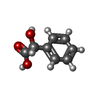
PDB-4p56: CRYSTAL STRUCTURE OF A TRAP PERIPLASMIC SOLUTE BINDING PROTEIN FROM BORDETELLA BRONCHISEPTICA, TARGET EFI-510038 (BB2442), WITH BOUND (R)-MANDELATE and (S)-MANDELATE Method: X-RAY DIFFRACTION / Resolution: 1.9 Å
PDB-4p8b: CRYSTAL STRUCTURE OF A TRAP PERIPLASMIC SOLUTE BINDING PROTEIN FROM RALSTONIA EUTROPHA H16 (H16_A1328), TARGET EFI-510189, WITH BOUND (S)-2-hydroxy-2-methyl-3-oxobutanoate ((S)-2-Acetolactate) Method: X-RAY DIFFRACTION / Resolution: 1.3 Å
PDB-4p9k: CRYSTAL STRUCTURE OF A TRAP PERIPLASMIC SOLUTE BINDING PROTEIN FROM VERMINEPHROBACTER EISENIAE EF01-2 (Veis_3954, TARGET EFI-510324) A NEPHRIDIAL SYMBIONT OF THE EARTHWORM EISENIA FOETIDA, BOUND TO D-ERYTHRONATE WITH RESIDUAL DENSITY SUGGESTIVE OF SUPERPOSITION WITH COPURIFIED ALTERNATIVE LIGAND. Method: X-RAY DIFFRACTION / Resolution: 1.4 Å
PDB-4paf: CRYSTAL STRUCTURE OF A TRAP PERIPLASMIC SOLUTE BINDING PROTEIN FROM RUEGERIA POMEROYI DSS-3 (SPO1773, TARGET EFI-510260) WITH BOUND 3,4-DIHYDROXYBENZOATE Method: X-RAY DIFFRACTION / Resolution: 1.6 Å
PDB-4pai: CRYSTAL STRUCTURE OF A TRAP PERIPLASMIC SOLUTE BINDING PROTEIN FROM RUEGERIA POMEROYI DSS-3 (SPO1773, TARGET EFI-510260) WITH BOUND 3-HYDROXYBENZOATE Method: X-RAY DIFFRACTION / Resolution: 1.4 Å
PDB-4pak: CRYSTAL STRUCTURE OF A TRAP PERIPLASMIC SOLUTE BINDING PROTEIN FROM VERMINEPHROBACTER EISENIAE EF01-2 (Veis_3954, TARGET EFI-510324) A NEPHRIDIAL SYMBIONT OF THE EARTHWORM EISENIA FOETIDA, BOUND TO (R)-PANTOIC ACID Method: X-RAY DIFFRACTION / Resolution: 1.2 Å
PDB-4pbh: CRYSTAL STRUCTURE OF A TRAP PERIPLASMIC SOLUTE BINDING PROTEIN FROM RUEGERIA POMEROYI DSS-3 (SPO1773, TARGET EFI-510260) WITH BOUND BENZOIC ACID Method: X-RAY DIFFRACTION / Resolution: 1.2 Å
PDB-4pbq: CRYSTAL STRUCTURE OF A TRAP PERIPLASMIC SOLUTE BINDING PROTEIN FROM HAEMOPHILUS INFLUENZAE RdAW (HICG_00826, TARGET EFI-510123) WITH BOUND L-GULONATE Method: X-RAY DIFFRACTION / Resolution: 1.65 Å
PDB-4pc9: CRYSTAL STRUCTURE OF A TRAP PERIPLASMIC SOLUTE BINDING PROTEIN FROM ROSENBACTER DENITRIFICANS OCh 114 (RD1_1052, TARGET EFI-510238) WITH BOUND D-MANNONATE Method: X-RAY DIFFRACTION / Resolution: 1.3 Å
PDB-4pcd: CRYSTAL STRUCTURE OF A TRAP PERIPLASMIC SOLUTE BINDING PROTEIN FROM ROSEOBACTER DENITRIFICANS OCh 114 (RD1_1052, TARGET EFI-510238) WITH BOUND L-GALACTONATE Method: X-RAY DIFFRACTION / Resolution: 1.7 Å
PDB-4pdd: CRYSTAL STRUCTURE OF A TRAP PERIPLASMIC SOLUTE BINDING PROTEIN FROM POLAROMONAS SP JS666 (Bpro_0088, TARGET EFI-510167) BOUND TO D-ERYTHRONATE Method: X-RAY DIFFRACTION / Resolution: 1.7 Å
PDB-4pdh: CRYSTAL STRUCTURE OF A TRAP PERIPLASMIC SOLUTE BINDING PROTEIN FROM POLAROMONAS SP JS666 (Bpro_1871, TARGET EFI-510164) BOUND TO D-ERYTHRONATE Method: X-RAY DIFFRACTION / Resolution: 1.8 Å
PDB-4pe3: CRYSTAL STRUCTURE OF A TRAP PERIPLASMIC SOLUTE BINDING PROTEIN FROM RHODOBACTER SPHAEROIDES (Rsph17029_3620, TARGET EFI-510199), APO OPEN STRUCTURE Method: X-RAY DIFFRACTION / Resolution: 1.35 Å
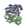
PDB-4pet: CRYSTAL STRUCTURE OF A TRAP PERIPLASMIC SOLUTE BINDING PROTEIN FROM COLWELLIA PSYCHRERYTHRAEA (CPS_0129, TARGET EFI-510097) WITH BOUND CALCIUM AND PYRUVATE Method: X-RAY DIFFRACTION / Resolution: 1.9 Å
PDB-4pf6: CRYSTAL STRUCTURE OF A TRAP PERIPLASMIC SOLUTE BINDING PROTEIN FROM ROSEOBACTER DENITRIFICANS (RD1_0742, TARGET EFI-510239) WITH BOUND 3-DEOXY-D-MANNO-OCT-2-ULOSONIC ACID (KDO) Method: X-RAY DIFFRACTION / Resolution: 1.75 Å
PDB-4pf8: CRYSTAL STRUCTURE OF A TRAP PERIPLASMIC SOLUTE BINDING PROTEIN FROM SULFITOBACTER sp. NAS-14.1 (TARGET EFI-510299) WITH BOUND BETA-D-GALACTURONATE Method: X-RAY DIFFRACTION / Resolution: 1.5 Å
PDB-4pfb: Crystal structure of a TRAP periplasmic solute binding protein from Fusobacterium nucleatun (FN1258, TARGET EFI-510120) with bound SN-glycerol-3-phosphate Method: X-RAY DIFFRACTION / Resolution: 2.7 Å
PDB-4pfi: Crystal structure of a tRAP periplasmic solute binding protein from marinobacter aquaeolei VT8 (Maqu_2829, TARGET EFI-510133), apo open structure Method: X-RAY DIFFRACTION / Resolution: 2.3024 Å
PDB-4pfr: CRYSTAL STRUCTURE OF A TRAP PERIPLASMIC SOLUTE BINDING PROTEIN FROM RHODOBACTER SPHAEROIDES (Rsph17029_3541, TARGET EFI-510203), APO OPEN PARTIALLY DISORDERED Method: X-RAY DIFFRACTION / Resolution: 2.6 Å
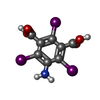
PDB-4pgn: CRYSTAL STRUCTURE OF A TRAP PERIPLASMIC SOLUTE BINDING PROTEIN FROM DESULFOVIBRIO ALASKENSIS G20 (Dde_0634, TARGET EFI-510120) WITH BOUND INDOLE PYRUVATE Method: X-RAY DIFFRACTION / Resolution: 1.8 Å
PDB-4pgp: CRYSTAL STRUCTURE OF A TRAP PERIPLASMIC SOLUTE BINDING PROTEIN FROM DESULFOVIBRIO ALASKENSIS G20 (Dde_0634, TARGET EFI-510120) WITH BOUND 3-INDOLE ACETIC ACID Method: X-RAY DIFFRACTION / Resolution: 2.25 Å
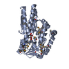
PDB-4uab: Crystal structure of a TRAP periplasmic solute binding protein from Chromohalobacter salexigens DSM 3043 (Csal_0678), Target EFI-501078, with bound ethanolamine Method: X-RAY DIFFRACTION / Resolution: 1.4 Å
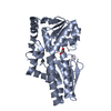
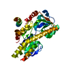
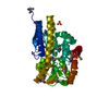
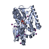
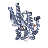
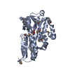
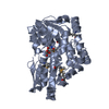
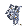
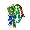
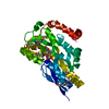
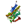
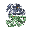
TRANSPORT PROTEIN / ENZYME FUNCTION INITIATIVE / EFI / Structural Genomics / TRAP periplasmic solute binding family / ABC PERIPLASMIC SOLUTE BINDING FAMILY / PROTEIN TRANSPORT / PROTEIN BINDING / MEMBRANE PROTEIN/PROTEIN TRANSPORT / MEMBRANE PROTEIN-PROTEIN TRANSPORT complex / SOLUTE-BINDING PROTEIN
+
About Yorodumi Papers
-
News
-
Feb 9, 2022. New format data for meta-information of EMDB entries
New format data for meta-information of EMDB entries
Version 3 of the EMDB header file is now the official format.
The previous official version 1.9 will be removed from the archive.
In the structure databanks used in Yorodumi, some data are registered as the other names, "COVID-19 virus" and "2019-nCoV". Here are the details of the virus and the list of structure data.
Jan 31, 2019. EMDB accession codes are about to change! (news from PDBe EMDB page)
EMDB accession codes are about to change! (news from PDBe EMDB page)
The allocation of 4 digits for EMDB accession codes will soon come to an end. Whilst these codes will remain in use, new EMDB accession codes will include an additional digit and will expand incrementally as the available range of codes is exhausted. The current 4-digit format prefixed with “EMD-” (i.e. EMD-XXXX) will advance to a 5-digit format (i.e. EMD-XXXXX), and so on. It is currently estimated that the 4-digit codes will be depleted around Spring 2019, at which point the 5-digit format will come into force.
The EM Navigator/Yorodumi systems omit the EMD- prefix.
Related info.:Q: What is EMD? / ID/Accession-code notation in Yorodumi/EM Navigator
 Movie
Movie Controller
 Structure viewers
 About Yorodumi Papers


 Authors
 External links
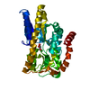


 Keywords
 burkholderia ambifaria (bacteria)
 pseudomonas aeruginosa (bacteria)