 Movie
Movie Controller

[English] 日本語
 Yorodumi
- PDB-4juo: A low-resolution three-gate structure of topoisomerase IV from St... -
+ Open data

- Basic information
| Entry | Database: PDB / ID: 4juo | ||||||
|---|---|---|---|---|---|---|---|
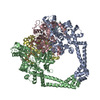
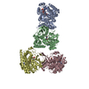
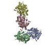
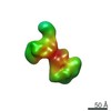
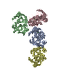
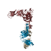
| Title | A low-resolution three-gate structure of topoisomerase IV from Streptococcus pneumoniae in space group H32 | ||||||
 Components Components |
| ||||||
 Keywords Keywords | ISOMERASE/DNA / full-length ParE / ParC55 / open N-gate / topoisomerase IIa / ATP binding / ISOMERASE-DNA complex | ||||||
| Function / homology |  Function and homology information Function and homology informationDNA topoisomerase type II (double strand cut, ATP-hydrolyzing) complex / DNA negative supercoiling activity / DNA topoisomerase (ATP-hydrolysing) / extrinsic component of plasma membrane / DNA topological change / chromosome segregation / chromosome / DNA binding / ATP binding / metal ion binding / cytoplasm Similarity search - Function | ||||||
| Biological species |   Streptococcus pneumoniae (bacteria)Streptococcus pneumoniae serotype 4 (bacteria) Streptococcus pneumoniae (bacteria)Streptococcus pneumoniae serotype 4 (bacteria) | ||||||
| Method |  X-RAY DIFFRACTION / SYNCHROTRON / MOLECULAR REPLACEMENT / Resolution: 6.53 Å X-RAY DIFFRACTION / SYNCHROTRON / MOLECULAR REPLACEMENT / Resolution: 6.53 Å | ||||||
 Authors Authors | Laponogov, I. / Veselkov, D.A. / Pan, X.-S. / Crevel, I. / Fisher, L.M. / Sanderson, M.R. | ||||||
 Citation Citation | Journal: Nucleic Acids Res. / Year: 2013 Title: Structure of an 'open' clamp type II topoisomerase-DNA complex provides a mechanism for DNA capture and transport. Authors: Laponogov, I. / Veselkov, D.A. / Crevel, I.M. / Pan, X.S. / Fisher, L.M. / Sanderson, M.R. | ||||||
| History |
|
- Structure visualization
| Structure viewer | Molecule: MolmilJmol/JSmol |
|---|
- Downloads & links
-Download
| PDBx/mmCIF format | 4juo.cif.gz | 432 KB | Display | PDBx/mmCIF format |
|---|---|---|---|---|
| PDB format | pdb4juo.ent.gz | 350.1 KB | Display | PDB format |
| PDBx/mmJSON format | 4juo.json.gz | Tree view | PDBx/mmJSON format | |
| Others |  Other downloads Other downloads |
-Validation report
| Arichive directory | https://data.pdbj.org/pub/pdb/validation_reports/ju/4juoftp://data.pdbj.org/pub/pdb/validation_reports/ju/4juo | HTTPS FTP |
|---|
-Related structure data
| Related structure data |  4i3hC  3raeS C: citing same article ( S: Starting model for refinement |
|---|---|
| Similar structure data |


-Links
 PDBj
PDBj


- Assembly
| Deposited unit | 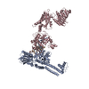
| ||||||||
|---|---|---|---|---|---|---|---|---|---|
| 1 |
| ||||||||
| Unit cell |
|
-Components
-DNA topoisomerase 4 subunit ... , 2 types, 2 molecules AC
| #1: Protein | Mass: 56455.434 Da / Num. of mol.: 1 / Fragment: ParC55 Source method: isolated from a genetically manipulated source Source: (gene. exp.) Streptococcus pneumoniae (bacteria) / Strain: ATCC BAA-334 / TIGR4 / Gene: parC, SP_0855 / Production host: |
|---|---|
| #2: Protein | Mass: 74528.180 Da / Num. of mol.: 1 / Fragment: ParE Source method: isolated from a genetically manipulated source Source: (gene. exp.) Streptococcus pneumoniae serotype 4 (bacteria)Strain: ATCC BAA-334 / TIGR4 / Gene: parE / Production host: |
-DNA chain , 4 types, 4 molecules EFGH
| #3: DNA chain | Mass: 3397.247 Da / Num. of mol.: 1 / Source method: obtained synthetically |
|---|---|
| #4: DNA chain | Mass: 4543.972 Da / Num. of mol.: 1 / Source method: obtained synthetically |
| #5: DNA chain | Mass: 3339.195 Da / Num. of mol.: 1 / Source method: obtained synthetically |
| #6: DNA chain | Mass: 4602.025 Da / Num. of mol.: 1 / Source method: obtained synthetically |
-Non-polymers , 2 types, 3 molecules 
| #7: Chemical |  Mass: 24.305 Da / Num. of mol.: 2 / Source method: obtained synthetically / Formula: Mg Mass: 24.305 Da / Num. of mol.: 2 / Source method: obtained synthetically / Formula: Mg#8: Chemical | ChemComp-LFX / ( |  Mass: 361.368 Da / Num. of mol.: 1 / Source method: obtained synthetically / Formula: C18H20FN3O4 Mass: 361.368 Da / Num. of mol.: 1 / Source method: obtained synthetically / Formula: C18H20FN3O4 |
|---|
-Experimental details
-Experiment
| Experiment | Method: X-RAY DIFFRACTION / Number of used crystals: 1 |
|---|
- Sample preparation
| Crystal | Density Matthews: 3.17 Å3/Da / Density % sol: 61.14 % |
|---|---|
| Crystal grow | Temperature: 293 K / Method: vapor diffusion, sitting drop / pH: 6.5 Details: 3-4% isopropanol, 50 mM Na cacodylate, optimised mixture of salts, pH 6.5, VAPOR DIFFUSION, SITTING DROP, temperature 293K |
-Data collection
| Diffraction | Mean temperature: 100 K |
|---|---|
| Diffraction source | Source: SYNCHROTRON / Site: Diamond  / Beamline: I03 / Wavelength: 0.9763 Å / Beamline: I03 / Wavelength: 0.9763 Å |
| Detector | Type: ADSC QUANTUM 315r / Detector: CCD / Date: Sep 26, 2010 |
| Radiation | Monochromator: Mirrors / Protocol: SINGLE WAVELENGTH / Monochromatic (M) / Laue (L): M / Scattering type: x-ray |
| Radiation wavelength | Wavelength: 0.9763 Å / Relative weight: 1 |
| Reflection | Resolution: 6.53→53.4 Å / Num. all: 3718 / Num. obs: 3701 / % possible obs: 99.24 % / Observed criterion σ(F): -1 / Observed criterion σ(I): -1 / Redundancy: 9.66 % / Rmerge(I) obs: 0.06 / Net I/σ(I): 6.84 |
| Reflection shell | Resolution: 6.53→6.71 Å / Redundancy: 10.93 % / Rmerge(I) obs: 0.41 / Mean I/σ(I) obs: 1.88 / Num. unique all: 309 / % possible all: 99.66 |
- Processing
| Software |
| |||||||||||||||||||||||||||||||||||||||||||||||||||||||||||||||||||||||||||||||||||||||||||||||||||||||||||||||||||||||||||||||||||||||||||||||||||||||||||||||||||||||||||||||||||||||||||||||||||||||||||||||||||||||||||||||||||||||||||||||||||||||||||||||||||||||||||||||||||||||||||||||||||||||||||||||||||||||||||||||||||||||||||||||||||||||||||||||||||||||||||||||||||||||
|---|---|---|---|---|---|---|---|---|---|---|---|---|---|---|---|---|---|---|---|---|---|---|---|---|---|---|---|---|---|---|---|---|---|---|---|---|---|---|---|---|---|---|---|---|---|---|---|---|---|---|---|---|---|---|---|---|---|---|---|---|---|---|---|---|---|---|---|---|---|---|---|---|---|---|---|---|---|---|---|---|---|---|---|---|---|---|---|---|---|---|---|---|---|---|---|---|---|---|---|---|---|---|---|---|---|---|---|---|---|---|---|---|---|---|---|---|---|---|---|---|---|---|---|---|---|---|---|---|---|---|---|---|---|---|---|---|---|---|---|---|---|---|---|---|---|---|---|---|---|---|---|---|---|---|---|---|---|---|---|---|---|---|---|---|---|---|---|---|---|---|---|---|---|---|---|---|---|---|---|---|---|---|---|---|---|---|---|---|---|---|---|---|---|---|---|---|---|---|---|---|---|---|---|---|---|---|---|---|---|---|---|---|---|---|---|---|---|---|---|---|---|---|---|---|---|---|---|---|---|---|---|---|---|---|---|---|---|---|---|---|---|---|---|---|---|---|---|---|---|---|---|---|---|---|---|---|---|---|---|---|---|---|---|---|---|---|---|---|---|---|---|---|---|---|---|---|---|---|---|---|---|---|---|---|---|---|---|---|---|---|---|---|---|---|---|---|---|---|---|---|---|---|---|---|---|---|---|---|---|---|---|---|---|---|---|---|---|---|---|---|---|---|---|---|---|---|---|---|---|---|---|---|---|---|---|---|---|---|---|---|---|---|---|---|---|---|---|---|---|---|---|---|---|---|---|---|---|---|---|---|---|---|---|---|---|---|---|---|---|---|---|---|---|---|---|---|
| Refinement | Method to determine structure: MOLECULAR REPLACEMENT Starting model: PDB ENTRY 3RAE Resolution: 6.53→46.442 Å / SU ML: 0.88 / Cross valid method: THROUGHOUT / σ(F): 1.36 / Phase error: 40.82 / Stereochemistry target values: ML Details: Refinement has been limited to rigid body and TLS refinement only due to the low resolution
| |||||||||||||||||||||||||||||||||||||||||||||||||||||||||||||||||||||||||||||||||||||||||||||||||||||||||||||||||||||||||||||||||||||||||||||||||||||||||||||||||||||||||||||||||||||||||||||||||||||||||||||||||||||||||||||||||||||||||||||||||||||||||||||||||||||||||||||||||||||||||||||||||||||||||||||||||||||||||||||||||||||||||||||||||||||||||||||||||||||||||||||||||||||||
| Solvent computation | Shrinkage radii: 0.9 Å / VDW probe radii: 1.11 Å / Solvent model: FLAT BULK SOLVENT MODEL | |||||||||||||||||||||||||||||||||||||||||||||||||||||||||||||||||||||||||||||||||||||||||||||||||||||||||||||||||||||||||||||||||||||||||||||||||||||||||||||||||||||||||||||||||||||||||||||||||||||||||||||||||||||||||||||||||||||||||||||||||||||||||||||||||||||||||||||||||||||||||||||||||||||||||||||||||||||||||||||||||||||||||||||||||||||||||||||||||||||||||||||||||||||||
| Refinement step | Cycle: LAST / Resolution: 6.53→46.442 Å
| |||||||||||||||||||||||||||||||||||||||||||||||||||||||||||||||||||||||||||||||||||||||||||||||||||||||||||||||||||||||||||||||||||||||||||||||||||||||||||||||||||||||||||||||||||||||||||||||||||||||||||||||||||||||||||||||||||||||||||||||||||||||||||||||||||||||||||||||||||||||||||||||||||||||||||||||||||||||||||||||||||||||||||||||||||||||||||||||||||||||||||||||||||||||
| Refine LS restraints |
| |||||||||||||||||||||||||||||||||||||||||||||||||||||||||||||||||||||||||||||||||||||||||||||||||||||||||||||||||||||||||||||||||||||||||||||||||||||||||||||||||||||||||||||||||||||||||||||||||||||||||||||||||||||||||||||||||||||||||||||||||||||||||||||||||||||||||||||||||||||||||||||||||||||||||||||||||||||||||||||||||||||||||||||||||||||||||||||||||||||||||||||||||||||||
| Refinement TLS params. | Method: refined / Refine-ID: X-RAY DIFFRACTION
| |||||||||||||||||||||||||||||||||||||||||||||||||||||||||||||||||||||||||||||||||||||||||||||||||||||||||||||||||||||||||||||||||||||||||||||||||||||||||||||||||||||||||||||||||||||||||||||||||||||||||||||||||||||||||||||||||||||||||||||||||||||||||||||||||||||||||||||||||||||||||||||||||||||||||||||||||||||||||||||||||||||||||||||||||||||||||||||||||||||||||||||||||||||||
| Refinement TLS group |
|
