 Movie
Movie Controller

[English] 日本語
 Yorodumi
- PDB-2nov: Breakage-reunion domain of S.pneumoniae topo IV: crystal structur... -
+ Open data

- Basic information
| Entry | Database: PDB / ID: 2nov | ||||||
|---|---|---|---|---|---|---|---|
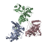
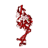
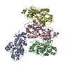
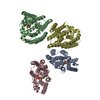
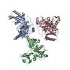
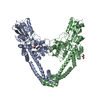
| Title | Breakage-reunion domain of S.pneumoniae topo IV: crystal structure of a gram-positive quinolone target | ||||||
 Components Components | DNA topoisomerase 4 subunit A | ||||||
 Keywords Keywords | ISOMERASE / protein / ParC / topo IV / gram-positive bacteria / quinolone target / DNA binding / DNA cleavage | ||||||
| Function / homology |  Function and homology information Function and homology informationDNA topoisomerase type II (double strand cut, ATP-hydrolyzing) complex / DNA negative supercoiling activity / DNA topoisomerase (ATP-hydrolysing) / extrinsic component of plasma membrane / DNA topological change / chromosome segregation / chromosome / DNA binding / ATP binding / cytoplasm Similarity search - Function | ||||||
| Biological species |   Streptococcus pneumoniae (bacteria) Streptococcus pneumoniae (bacteria) | ||||||
| Method |  X-RAY DIFFRACTION / SYNCHROTRON / MOLECULAR REPLACEMENT / Resolution: 2.67 Å X-RAY DIFFRACTION / SYNCHROTRON / MOLECULAR REPLACEMENT / Resolution: 2.67 Å | ||||||
 Authors Authors | Laponogov, I. / Veselkov, D.A. / Sohi, M.K. / Pan, X.S. / Achari, A. / Yang, C. / Ferrara, J.D. / Fisher, L.M. / Sanderson, M.R. | ||||||
 Citation Citation | Journal: PLoS ONE / Year: 2007 Title: Breakage-Reunion Domain of Streptococcus pneumoniae Topoisomerase IV: Crystal Structure of a Gram-Positive Quinolone Target. Authors: Laponogov, I. / Veselkov, D.A. / Sohi, M.K. / Pan, X.S. / Achari, A. / Yang, C. / Ferrara, J.D. / Fisher, L.M. / Sanderson, M.R. #1: Journal: Acta Crystallogr.,Sect.A / Year: 2005Title: The Structure of the ParC Subunit of Topoisomerase IV from Streptococcus pneumoniae Authors: Laponogov, I. / Veselkov, D.A. / Sohi, M.K. / Pan, X.S. / Fisher, L.M. / Sanderson, M.R. | ||||||
| History |
|
- Structure visualization
| Structure viewer | Molecule: MolmilJmol/JSmol |
|---|
- Downloads & links
-Download
| PDBx/mmCIF format | 2nov.cif.gz | 341.6 KB | Display | PDBx/mmCIF format |
|---|---|---|---|---|
| PDB format | pdb2nov.ent.gz | 277.4 KB | Display | PDB format |
| PDBx/mmJSON format | 2nov.json.gz | Tree view | PDBx/mmJSON format | |
| Others |  Other downloads Other downloads |
-Validation report
| Arichive directory | https://data.pdbj.org/pub/pdb/validation_reports/no/2novftp://data.pdbj.org/pub/pdb/validation_reports/no/2nov | HTTPS FTP |
|---|
-Related structure data
| Related structure data |  1ab4S S: Starting model for refinement |
|---|---|
| Similar structure data |


-Links
 PDBj
PDBj

- Assembly
| Deposited unit | 
| ||||||||
|---|---|---|---|---|---|---|---|---|---|
| 1 | 
| ||||||||
| 2 | 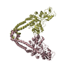
| ||||||||
| Unit cell |
| ||||||||
| Details | The biological assembly is a dimer generated by a) A and B chains b) C and D chains (two biological dimers in the asymmetric unit) |
-Components
| #1: Protein | Mass: 56455.434 Da / Num. of mol.: 4 / Fragment: 55-kDa N-terminal fragment Source method: isolated from a genetically manipulated source Source: (gene. exp.) Streptococcus pneumoniae (bacteria) / Strain: 7785 / Gene: parC / Plasmid: pET29a / Production host: References: UniProt: P72525, UniProt: Q3HYJ3*PLUS, Isomerases; Other isomerases; Sole sub-subclass for isomerases that do not belong in the other subclasses #2: Water | ChemComp-HOH / |  Mass: 18.015 Da / Num. of mol.: 24 / Source method: isolated from a natural source / Formula: H2O Mass: 18.015 Da / Num. of mol.: 24 / Source method: isolated from a natural source / Formula: H2O |
|---|
-Experimental details
-Experiment
| Experiment | Method: X-RAY DIFFRACTION / Number of used crystals: 1 |
|---|
- Sample preparation
| Crystal | Density Matthews: 3.4 Å3/Da / Density % sol: 63.88 % |
|---|---|
| Crystal grow | Temperature: 298 K / Method: vapor diffusion, hanging drop / pH: 5.5 Details: The purified ParC55 protein was dialyzed against 20 mM Tris-HCl, pH 7.0, 200 mM NaCl, 10% glycerol, 1 mM beta-mercaptoethanol, 0.05% NaN3 then concentrated to 3-4 mg/ml. Hanging drops were ...Details: The purified ParC55 protein was dialyzed against 20 mM Tris-HCl, pH 7.0, 200 mM NaCl, 10% glycerol, 1 mM beta-mercaptoethanol, 0.05% NaN3 then concentrated to 3-4 mg/ml. Hanging drops were formed by mixing equal volumes of protein and crystallization solutions (100 mM Tris-HCl, pH 5.5, 200 mM NaCl, 1 mM beta-mercaptoethanol, 0.05% NaN3 and 10% of 1:1 ethanol-isopropanol as precipitant). , VAPOR DIFFUSION, HANGING DROP, temperature 298.0K |
-Data collection
| Diffraction | Mean temperature: 100 K |
|---|---|
| Diffraction source | Source: SYNCHROTRON / Site: ESRF  / Beamline: BM30A / Wavelength: 0.91694 Å / Beamline: BM30A / Wavelength: 0.91694 Å |
| Detector | Type: MAR scanner 345 mm plate / Detector: IMAGE PLATE / Details: two cylindrical parabolic mirrors |
| Radiation | Protocol: SINGLE WAVELENGTH / Monochromatic (M) / Laue (L): M / Scattering type: x-ray |
| Radiation wavelength | Wavelength: 0.91694 Å / Relative weight: 1 |
| Reflection twin | Type: hemihedral / Operator: k,-h,l / Fraction: 0.323 |
| Reflection | Highest resolution: 2.67 Å / Num. obs: 85197 / Redundancy: 5.4 % / Rsym value: 0.093 |
- Processing
| Software |
| ||||||||||||||||||||||||||||
|---|---|---|---|---|---|---|---|---|---|---|---|---|---|---|---|---|---|---|---|---|---|---|---|---|---|---|---|---|---|
| Refinement | Method to determine structure: MOLECULAR REPLACEMENT Starting model: PDB ENTRY 1AB4 Resolution: 2.67→500 Å Details: Refinement was performed using twinning operator (k,-h,l) and twinning fraction of 0.323
| ||||||||||||||||||||||||||||
| Solvent computation | Bsol: 13.223 Å2 | ||||||||||||||||||||||||||||
| Displacement parameters | Biso mean: 64.915 Å2
| ||||||||||||||||||||||||||||
| Refine analyze |
| ||||||||||||||||||||||||||||
| Refinement step | Cycle: LAST / Resolution: 2.67→500 Å
| ||||||||||||||||||||||||||||
| Refine LS restraints |
| ||||||||||||||||||||||||||||
| Xplor file |
|
