 Movie
Movie Controller


+ Open data

- Basic information
| Entry | Database: PDB / ID: 1i7o | ||||||
|---|---|---|---|---|---|---|---|
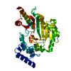
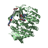
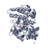
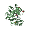
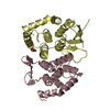
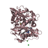
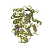
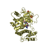
| Title | CRYSTAL STRUCTURE OF HPCE | ||||||
 Components Components | 4-HYDROXYPHENYLACETATE DEGRADATION BIFUNCTIONAL ISOMERASE/DECARBOXYLASE | ||||||
 Keywords Keywords | ISOMERASE / LYASE / bifunctional enzyme / decarboxylase | ||||||
| Function / homology |  Function and homology information Function and homology information5-oxopent-3-ene-1,2,5-tricarboxylate decarboxylase / 5-oxopent-3-ene-1,2,5-tricarboxylate decarboxylase activity / 5-carboxymethyl-2-hydroxymuconate Delta-isomerase / 4-hydroxyphenylacetate catabolic process / 5-carboxymethyl-2-hydroxymuconate delta-isomerase activity / metal ion binding Similarity search - Function | ||||||
| Biological species |  | ||||||
| Method |  X-RAY DIFFRACTION / SYNCHROTRON / MIR / Resolution: 1.7 Å X-RAY DIFFRACTION / SYNCHROTRON / MIR / Resolution: 1.7 Å | ||||||
 Authors Authors | Tame, J.R.H. / Namba, K. / Dodson, E.J. / Roper, D.I. | ||||||
 Citation Citation | Journal: To be Published Title: The crystal structure of HpcE, a multi-functional enzyme fold Authors: Tame, J.R.H. / Namba, K. / Dodson, E.J. / Roper, D.I. | ||||||
| History |
|
- Structure visualization
| Structure viewer | Molecule: MolmilJmol/JSmol |
|---|
- Downloads & links
-Download
| PDBx/mmCIF format | 1i7o.cif.gz | 339.3 KB | Display | PDBx/mmCIF format |
|---|---|---|---|---|
| PDB format | pdb1i7o.ent.gz | 275.1 KB | Display | PDB format |
| PDBx/mmJSON format | 1i7o.json.gz | Tree view | PDBx/mmJSON format | |
| Others |  Other downloads Other downloads |
-Validation report
| Arichive directory | https://data.pdbj.org/pub/pdb/validation_reports/i7/1i7oftp://data.pdbj.org/pub/pdb/validation_reports/i7/1i7o | HTTPS FTP |
|---|
-Related structure data
| Similar structure data |
|---|


-Links
 PDBj
PDBj- Assembly
| Deposited unit | 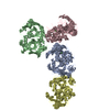
| ||||||||
|---|---|---|---|---|---|---|---|---|---|
| 1 | 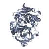
| ||||||||
| 2 | 
| ||||||||
| 3 | 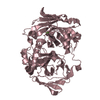
| ||||||||
| 4 | 
| ||||||||
| Unit cell |
|
-Components
| #1: Protein | Mass: 47154.156 Da / Num. of mol.: 4 Source method: isolated from a genetically manipulated source Source: (gene. exp.) References: UniProt: P37352, 5-carboxymethyl-2-hydroxymuconate Delta-isomerase, 5-oxopent-3-ene-1,2,5-tricarboxylate decarboxylase #2: Chemical | ChemComp-CA /   Mass: 40.078 Da / Num. of mol.: 4 / Source method: obtained synthetically / Formula: Ca Mass: 40.078 Da / Num. of mol.: 4 / Source method: obtained synthetically / Formula: Ca#3: Water | ChemComp-HOH / |  Mass: 18.015 Da / Num. of mol.: 668 / Source method: isolated from a natural source / Formula: H2O Mass: 18.015 Da / Num. of mol.: 668 / Source method: isolated from a natural source / Formula: H2OHas protein modification | Y | |
|---|
-Experimental details
-Experiment
| Experiment | Method: X-RAY DIFFRACTION / Number of used crystals: 1 |
|---|
- Sample preparation
| Crystal | Density Matthews: 2.4 Å3/Da / Density % sol: 48.74 % | ||||||||||||||||||||||||||||||
|---|---|---|---|---|---|---|---|---|---|---|---|---|---|---|---|---|---|---|---|---|---|---|---|---|---|---|---|---|---|---|---|
| Crystal grow | Temperature: 291 K / Method: vapor diffusion, hanging drop / pH: 8 Details: monomethylether PEG2000, calcium chloride, Tris, pH 8.0, VAPOR DIFFUSION, HANGING DROP, temperature 291K | ||||||||||||||||||||||||||||||
| Crystal grow | *PLUS Temperature: 20 ℃Details: drop contains protein and reservoir solution in a 1:1 ratio | ||||||||||||||||||||||||||||||
| Components of the solutions | *PLUS
|
-Data collection
| Diffraction source | Source: SYNCHROTRON / Site: EMBL/DESY, HAMBURG  / Beamline: X11 / Wavelength: 0.87 Å / Beamline: X11 / Wavelength: 0.87 Å |
|---|---|
| Detector | Type: MARRESEARCH / Detector: IMAGE PLATE / Date: Apr 4, 1999 |
| Radiation | Protocol: SINGLE WAVELENGTH / Monochromatic (M) / Laue (L): M / Scattering type: x-ray |
| Radiation wavelength | Wavelength: 0.87 Å / Relative weight: 1 |
| Reflection | Resolution: 1.7→15 Å / Num. all: 198751 / Num. obs: 196319 / % possible obs: 98.8 % / Observed criterion σ(F): 0 / Observed criterion σ(I): 0 / Redundancy: 3.4 % / Biso Wilson estimate: 22 Å2 / Rmerge(I) obs: 0.055 / Net I/σ(I): 18.6 |
| Reflection shell | Resolution: 1.7→1.75 Å / Redundancy: 3 % / Rmerge(I) obs: 0.546 / Mean I/σ(I) obs: 2.2 / % possible all: 97.5 |
| Reflection | *PLUS Lowest resolution: 91.2 Å / % possible obs: 98.5 % |
| Reflection shell | *PLUS % possible obs: 98.3 % |
- Processing
| Software |
| ||||||||||||||||||||||||||||||||||||||||||||||||||||||||||||||||||||||||||||||||||||
|---|---|---|---|---|---|---|---|---|---|---|---|---|---|---|---|---|---|---|---|---|---|---|---|---|---|---|---|---|---|---|---|---|---|---|---|---|---|---|---|---|---|---|---|---|---|---|---|---|---|---|---|---|---|---|---|---|---|---|---|---|---|---|---|---|---|---|---|---|---|---|---|---|---|---|---|---|---|---|---|---|---|---|---|---|---|
| Refinement | Method to determine structure: MIR / Resolution: 1.7→15 Å / Cross valid method: THROUGHOUT / σ(F): 0 / σ(I): 0 / Stereochemistry target values: Engh & Huber
| ||||||||||||||||||||||||||||||||||||||||||||||||||||||||||||||||||||||||||||||||||||
| Displacement parameters | Biso mean: 44.65 Å2
| ||||||||||||||||||||||||||||||||||||||||||||||||||||||||||||||||||||||||||||||||||||
| Refinement step | Cycle: LAST / Resolution: 1.7→15 Å
| ||||||||||||||||||||||||||||||||||||||||||||||||||||||||||||||||||||||||||||||||||||
| Refine LS restraints |
| ||||||||||||||||||||||||||||||||||||||||||||||||||||||||||||||||||||||||||||||||||||
| Software | *PLUS Name: REFMAC / Classification: refinement | ||||||||||||||||||||||||||||||||||||||||||||||||||||||||||||||||||||||||||||||||||||
| Refinement | *PLUS Lowest resolution: 91.2 Å / Num. reflection obs: 186453 / Rfactor Rfree: 0.252 / Rfactor Rwork: 0.2155 | ||||||||||||||||||||||||||||||||||||||||||||||||||||||||||||||||||||||||||||||||||||
| Solvent computation | *PLUS | ||||||||||||||||||||||||||||||||||||||||||||||||||||||||||||||||||||||||||||||||||||
| Displacement parameters | *PLUS | ||||||||||||||||||||||||||||||||||||||||||||||||||||||||||||||||||||||||||||||||||||
| Refine LS restraints | *PLUS
|
