Mass: 18.015 Da / Num. of mol.: 547 / Source method: isolated from a natural source / Formula: H2O
Sequence details
DISCREPANCIES IN SEQUENCE BETWEEN THE UNIPROT ENTRY Q004B5 AND THE SEQUENCE DETERMINED FROM ...DISCREPANCIES IN SEQUENCE BETWEEN THE UNIPROT ENTRY Q004B5 AND THE SEQUENCE DETERMINED FROM ELECTRON DENSITY ARE CLEARLY SUPPORTED BY THE ELECTRON DENSITY. RESIDUES K43, M131, M139, M201, I286 AND A295 ARE INDEED SEQUENCED BY THE ELECTRON DENSITY AT 1.25 A OF THIS DEPOSIT.
-
Experimental details
-
Experiment
Experiment
Method: X-RAY DIFFRACTION / Number of used crystals: 1
-
Sample preparation
Crystal
Density Matthews: 2.11 Å3/Da / Density % sol: 41.84 % / Description: NONE
Crystal grow
pH: 7.4 Details: 0.2 M SODIUM ACETATE, 0.1 M SODIUM CACODYLATE PH 6.5 AND 30% PEG 8000
Type: ADSC CCD / Detector: CCD / Date: Dec 12, 2012 Details: DOUBLE CRYSTAL CHANNEL CUT, SI(111), 1M LONG RH COATED TOROIDAL MIRROR FOR VERTICAL AND HORIZONTAL FOCUSING.
Radiation
Protocol: SINGLE WAVELENGTH / Monochromatic (M) / Laue (L): M / Scattering type: x-ray
Radiation wavelength
Wavelength: 0.9795 Å / Relative weight: 1
Reflection
Resolution: 1.25→30 Å / Num. obs: 91314 / % possible obs: 100 % / Observed criterion σ(I): 0 / Redundancy: 7.16 % / Biso Wilson estimate: 16.418 Å2 / Rmerge(I) obs: 0.05 / Net I/σ(I): 22.25
Reflection shell
Resolution: 1.25→1.28 Å / Redundancy: 7.1 % / Rmerge(I) obs: 0.54 / Mean I/σ(I) obs: 3.96 / % possible all: 100
-
Processing
Software
Name
Version
Classification
PHENIX
(PHENIX.REFINE)
refinement
XDS
datareduction
XSCALE
datascaling
PHASER
phasing
Refinement
Method to determine structure: MOLECULAR REPLACEMENT Starting model: A THEORETICAL MOLECULAR MODEL OF THE SHRIMP AMINO ACID SEQUENCE GENBANK ABI98020.1 BUILT WITH MOE CHEMCOMP AS A STARTING MODEL Resolution: 1.25→28.095 Å / SU ML: 0.34 / σ(F): 1.33 / Phase error: 20.36 / Stereochemistry target values: ML
Rfactor
Num. reflection
% reflection
Rfree
0.1968
4573
5 %
Rwork
0.1584
-
-
obs
0.1607
91313
99.96 %
Solvent computation
Shrinkage radii: 0.41 Å / VDW probe radii: 0.6 Å / Solvent model: FLAT BULK SOLVENT MODEL / Bsol: 56.338 Å2 / ksol: 0.461 e/Å3
Displacement parameters
Biso mean: 16.07 Å2
Baniso -1
Baniso -2
Baniso -3
1-
0.1012 Å2
0 Å2
0 Å2
2-
-
-0.0593 Å2
0 Å2
3-
-
-
-0.0419 Å2
Refinement step
Cycle: LAST / Resolution: 1.25→28.095 Å
Protein
Nucleic acid
Ligand
Solvent
Total
Num. atoms
2733
0
0
547
3280
Refine LS restraints
Refine-ID
Type
Dev ideal
Number
X-RAY DIFFRACTION
f_bond_d
0.012
2928
X-RAY DIFFRACTION
f_angle_d
1.339
3957
X-RAY DIFFRACTION
f_dihedral_angle_d
12.531
1124
X-RAY DIFFRACTION
f_chiral_restr
0.074
427
X-RAY DIFFRACTION
f_plane_restr
0.008
519
LS refinement shell
Resolution (Å)
Rfactor Rfree
Num. reflection Rfree
Rfactor Rwork
Num. reflection Rwork
Refine-ID
% reflection obs (%)
1.25-1.2642
0.348
143
0.2929
2870
X-RAY DIFFRACTION
100
1.2642-1.2791
0.3523
141
0.2898
2846
X-RAY DIFFRACTION
100
1.2791-1.2947
0.3593
161
0.2753
2878
X-RAY DIFFRACTION
100
1.2947-1.3111
0.3532
153
0.2718
2862
X-RAY DIFFRACTION
100
1.3111-1.3283
0.3451
132
0.2622
2865
X-RAY DIFFRACTION
100
1.3283-1.3465
0.3214
164
0.2496
2857
X-RAY DIFFRACTION
100
1.3465-1.3658
0.3059
139
0.2402
2846
X-RAY DIFFRACTION
100
1.3658-1.3861
0.302
169
0.2226
2885
X-RAY DIFFRACTION
100
1.3861-1.4078
0.2994
144
0.2166
2844
X-RAY DIFFRACTION
100
1.4078-1.4309
0.2689
134
0.1928
2874
X-RAY DIFFRACTION
100
1.4309-1.4556
0.2761
163
0.1837
2854
X-RAY DIFFRACTION
100
1.4556-1.482
0.2307
167
0.1781
2854
X-RAY DIFFRACTION
100
1.482-1.5105
0.2542
160
0.1657
2857
X-RAY DIFFRACTION
100
1.5105-1.5414
0.2309
135
0.1689
2881
X-RAY DIFFRACTION
100
1.5414-1.5749
0.2236
141
0.1573
2889
X-RAY DIFFRACTION
100
1.5749-1.6115
0.2075
154
0.151
2894
X-RAY DIFFRACTION
100
1.6115-1.6518
0.2171
126
0.1425
2881
X-RAY DIFFRACTION
100
1.6518-1.6964
0.2148
176
0.1406
2856
X-RAY DIFFRACTION
100
1.6964-1.7464
0.2263
169
0.1367
2867
X-RAY DIFFRACTION
100
1.7464-1.8027
0.2053
141
0.1363
2889
X-RAY DIFFRACTION
100
1.8027-1.8671
0.189
170
0.1397
2877
X-RAY DIFFRACTION
100
1.8671-1.9419
0.2084
157
0.1431
2888
X-RAY DIFFRACTION
100
1.9419-2.0302
0.1893
150
0.1367
2916
X-RAY DIFFRACTION
100
2.0302-2.1372
0.1926
148
0.1311
2897
X-RAY DIFFRACTION
100
2.1372-2.2711
0.1671
152
0.1268
2923
X-RAY DIFFRACTION
100
2.2711-2.4463
0.1831
155
0.1305
2906
X-RAY DIFFRACTION
100
2.4463-2.6923
0.1591
155
0.1425
2933
X-RAY DIFFRACTION
100
2.6923-3.0815
0.1829
152
0.1447
2949
X-RAY DIFFRACTION
100
3.0815-3.8808
0.1643
163
0.1427
2981
X-RAY DIFFRACTION
100
3.8808-28.1021
0.1764
159
0.1631
3121
X-RAY DIFFRACTION
100
+
About Yorodumi
-
News
-
Feb 9, 2022. New format data for meta-information of EMDB entries
New format data for meta-information of EMDB entries
Version 3 of the EMDB header file is now the official format.
The previous official version 1.9 will be removed from the archive.
In the structure databanks used in Yorodumi, some data are registered as the other names, "COVID-19 virus" and "2019-nCoV". Here are the details of the virus and the list of structure data.
Jan 31, 2019. EMDB accession codes are about to change! (news from PDBe EMDB page)
EMDB accession codes are about to change! (news from PDBe EMDB page)
The allocation of 4 digits for EMDB accession codes will soon come to an end. Whilst these codes will remain in use, new EMDB accession codes will include an additional digit and will expand incrementally as the available range of codes is exhausted. The current 4-digit format prefixed with “EMD-” (i.e. EMD-XXXX) will advance to a 5-digit format (i.e. EMD-XXXXX), and so on. It is currently estimated that the 4-digit codes will be depleted around Spring 2019, at which point the 5-digit format will come into force.
The EM Navigator/Yorodumi systems omit the EMD- prefix.
Related info.:Q: What is EMD? / ID/Accession-code notation in Yorodumi/EM Navigator
Yorodumi is a browser for structure data from EMDB, PDB, SASBDB, etc.
This page is also the successor to EM Navigator detail page, and also detail information page/front-end page for Omokage search.
The word "yorodu" (or yorozu) is an old Japanese word meaning "ten thousand". "mi" (miru) is to see.
Related info.:EMDB / PDB / SASBDB / Comparison of 3 databanks / Yorodumi Search / Aug 31, 2016. New EM Navigator & Yorodumi / Yorodumi Papers / Jmol/JSmol / Function and homology information / Changes in new EM Navigator and Yorodumi
 Movie
Movie Controller

 Yorodumi
 Open data
 Basic information
 Components
 Keywords
 Function and homology information
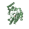
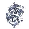
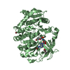
 LITOPENAEUS VANNAMEI (Pacific white shrimp)
 X-RAY DIFFRACTION /
  Authors
 Citation
 Structure visualization
 Downloads & links
 Other downloads
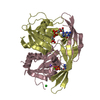


 PDBj
PDBj
 Assembly
 Mass: 18.015 Da / Num. of mol.: 547 / Source method: isolated from a natural source / Formula: H2O
 Sample preparation
 / Beamline: X6A / Wavelength: 0.9795
  Processing