 Movie
Movie Controller


+ Open data

- Basic information
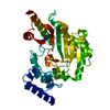
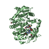
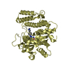
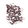
| Entry | Database: PDB / ID: 4xyi | ||||||
|---|---|---|---|---|---|---|---|
| Title | Mis16 with H4 peptide | ||||||
 Components Components |
| ||||||
 Keywords Keywords | CHAPERONE / Centromere / CENP-A / kinetochore / Mis18 complex / histone | ||||||
| Function / homology |  Function and homology information Function and homology informationCENP-A recruiting complex / Rpd3L complex / Rpd3L-Expanded complex / CENP-A containing chromatin assembly / kinetochore / structural constituent of chromatin / nucleosome / nucleosome assembly / histone binding / chromatin remodeling ...CENP-A recruiting complex / Rpd3L complex / Rpd3L-Expanded complex / CENP-A containing chromatin assembly / kinetochore / structural constituent of chromatin / nucleosome / nucleosome assembly / histone binding / chromatin remodeling / protein heterodimerization activity / regulation of DNA-templated transcription / DNA binding / nucleus / cytoplasm Similarity search - Function | ||||||
| Biological species | Schizosaccharomyces japonicus | ||||||
| Method |  X-RAY DIFFRACTION / SYNCHROTRON / MOLECULAR REPLACEMENT / Resolution: 3 Å X-RAY DIFFRACTION / SYNCHROTRON / MOLECULAR REPLACEMENT / Resolution: 3 Å | ||||||
 Authors Authors | An, S. / Kim, H. / Cho, U.-S. | ||||||
 Citation Citation | Journal: J.Mol.Biol. / Year: 2015 Title: Mis16 Independently Recognizes Histone H4 and the CENP-ACnp1-Specific Chaperone Scm3sp. Authors: An, S. / Kim, H. / Cho, U.S. | ||||||
| History |
|
- Structure visualization
| Structure viewer | Molecule: MolmilJmol/JSmol |
|---|
- Downloads & links
-Download
| PDBx/mmCIF format | 4xyi.cif.gz | 92.2 KB | Display | PDBx/mmCIF format |
|---|---|---|---|---|
| PDB format | pdb4xyi.ent.gz | 67.3 KB | Display | PDB format |
| PDBx/mmJSON format | 4xyi.json.gz | Tree view | PDBx/mmJSON format | |
| Others |  Other downloads Other downloads |
-Validation report
| Arichive directory | https://data.pdbj.org/pub/pdb/validation_reports/xy/4xyiftp://data.pdbj.org/pub/pdb/validation_reports/xy/4xyi | HTTPS FTP |
|---|
-Related structure data
| Related structure data | 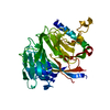 4xyhSC S: Starting model for refinement C: citing same article ( |
|---|---|
| Similar structure data |


-Links
 PDBj
PDBj


- Assembly
| Deposited unit | 
| ||||||||
|---|---|---|---|---|---|---|---|---|---|
| 1 |
| ||||||||
| Unit cell |
|
-Components
| #1: Protein | Mass: 48462.539 Da / Num. of mol.: 1 Source method: isolated from a genetically manipulated source Source: (gene. exp.)  Strain: yFS275 / FY16936 / Gene: SJAG_03867 / Plasmid: pFastBac HTb / Cell line (production host): Hi-5 / Production host:  Baculovirus expression vector pFastBac1-HM / References: UniProt: B6K598 Baculovirus expression vector pFastBac1-HM / References: UniProt: B6K598 |
|---|---|
| #2: Protein | Mass: 11420.441 Da / Num. of mol.: 1 / Source method: obtained synthetically Source: (synth.) References: UniProt: B6JV96 |
-Experimental details
-Experiment
| Experiment | Method: X-RAY DIFFRACTION / Number of used crystals: 1 |
|---|
- Sample preparation
| Crystal | Density Matthews: 2.94 Å3/Da / Density % sol: 58.23 % |
|---|---|
| Crystal grow | Temperature: 293 K / Method: vapor diffusion, hanging drop / pH: 7.5 Details: 0.5 M disodium phosphate 0.5 M dipotassium phosphate 0.1 M ammonium sulfate |
-Data collection
| Diffraction | Mean temperature: 105 K |
|---|---|
| Diffraction source | Source: SYNCHROTRON / Site: APS  / Beamline: 21-ID-G / Wavelength: 0.978 Å / Beamline: 21-ID-G / Wavelength: 0.978 Å |
| Detector | Type: MARMOSAIC 300 mm CCD / Detector: CCD / Date: Feb 20, 2014 |
| Radiation | Protocol: SINGLE WAVELENGTH / Monochromatic (M) / Laue (L): M / Scattering type: x-ray |
| Radiation wavelength | Wavelength: 0.978 Å / Relative weight: 1 |
| Reflection | Resolution: 3→50 Å / Num. obs: 14170 / % possible obs: 99.52 % / Redundancy: 8.8 % / Rsym value: 0.11 / Net I/σ(I): 15.7 |
| Reflection shell | Resolution: 3→3.11 Å / Mean I/σ(I) obs: 2.5 / % possible all: 98.8 |
- Processing
| Software |
| |||||||||||||||||||||||||||||||||||||||||||||||||||||||||||||||||||||||||||||
|---|---|---|---|---|---|---|---|---|---|---|---|---|---|---|---|---|---|---|---|---|---|---|---|---|---|---|---|---|---|---|---|---|---|---|---|---|---|---|---|---|---|---|---|---|---|---|---|---|---|---|---|---|---|---|---|---|---|---|---|---|---|---|---|---|---|---|---|---|---|---|---|---|---|---|---|---|---|---|
| Refinement | Method to determine structure: MOLECULAR REPLACEMENT Starting model: 4XYH Resolution: 3→39.325 Å / SU ML: 0.34 / Cross valid method: FREE R-VALUE / σ(F): 1.37 / Phase error: 29.87 / Stereochemistry target values: ML
| |||||||||||||||||||||||||||||||||||||||||||||||||||||||||||||||||||||||||||||
| Solvent computation | Shrinkage radii: 0.9 Å / VDW probe radii: 1.11 Å / Solvent model: FLAT BULK SOLVENT MODEL | |||||||||||||||||||||||||||||||||||||||||||||||||||||||||||||||||||||||||||||
| Refinement step | Cycle: LAST / Resolution: 3→39.325 Å
| |||||||||||||||||||||||||||||||||||||||||||||||||||||||||||||||||||||||||||||
| Refine LS restraints |
| |||||||||||||||||||||||||||||||||||||||||||||||||||||||||||||||||||||||||||||
| LS refinement shell |
|
