[English] 日本語
Yorodumi
- PDB-4x8b: Ergothioneine-biosynthetic sulfoxide synthase EgtB, apo form -

+
Open data


ID or keywords:

Loading...

-
Basic information

Entry
Database: PDB / ID: 4x8b
TitleErgothioneine-biosynthetic sulfoxide synthase EgtB, apo form
ComponentsSulfoxide synthase EgtB
KeywordsOXIDOREDUCTASE / ergothioneine biosynthesis / C-Lectin / DinB / non-heme Fe(II) enzyme
Function / homology
Function and homology information


gamma-glutamyl hercynylcysteine S-oxide synthase / gamma-glutamyl hercynylcysteine sulfoxide synthase activity / monooxygenase activity / iron ion binding
Similarity search - Function
Hercynine oxygenase, Actinobacteria / Ergothioneine biosynthesis protein EgtB / DinB-like domain / DinB superfamily / DinB/YfiT-like putative metalloenzymes / Sulfatase-modifying factor enzyme / Sulfatase-modifying factor enzyme 1 / Sulfatase-modifying factor enzyme superfamily / C-type lectin fold
Similarity search - Domain/homology
: / Hercynine oxygenase
Similarity search - Component
Biological speciesMycobacterium thermoresistibile ATCC 19527 (bacteria)
MethodX-RAY DIFFRACTION / SYNCHROTRON / SAD / Resolution: 1.7 Å
AuthorsVit, A. / Goncharenko, K.V. / Blankenfeldt, W. / Seebeck, F.P.
Funding support Switzerland, 1items
OrganizationGrant numberCountry
Swiss National Science Foundation1147005 Switzerland
CitationJournal: Angew.Chem.Int.Ed.Engl. / Year: 2015
Title: Structure of the Sulfoxide Synthase EgtB from the Ergothioneine Biosynthetic Pathway.
Authors: Goncharenko, K.V. / Vit, A. / Blankenfeldt, W. / Seebeck, F.P.
History
DepositionDec 10, 2014Deposition site: RCSB / Processing site: PDBE
Revision 1.0Jan 28, 2015Provider: repository / Type: Initial release
Revision 1.1Feb 25, 2015Group: Database references
Revision 2.0Sep 6, 2017Group: Advisory / Atomic model ...Advisory / Atomic model / Author supporting evidence / Data collection / Derived calculations / Refinement description
Category: atom_site / diffrn_radiation_wavelength ...atom_site / diffrn_radiation_wavelength / pdbx_audit_support / pdbx_distant_solvent_atoms / pdbx_struct_conn_angle / pdbx_validate_close_contact / software / struct_conn / struct_site_gen
Item: _atom_site.B_iso_or_equiv / _atom_site.Cartn_x ..._atom_site.B_iso_or_equiv / _atom_site.Cartn_x / _atom_site.Cartn_y / _atom_site.Cartn_z / _pdbx_audit_support.funding_organization / _pdbx_distant_solvent_atoms.auth_seq_id / _pdbx_struct_conn_angle.ptnr1_auth_seq_id / _pdbx_struct_conn_angle.ptnr3_auth_seq_id / _pdbx_struct_conn_angle.value / _pdbx_validate_close_contact.auth_seq_id_1 / _pdbx_validate_close_contact.auth_seq_id_2 / _software.name / _struct_conn.pdbx_dist_value / _struct_conn.ptnr2_auth_seq_id / _struct_site_gen.auth_seq_id

-
Structure visualization

Structure viewerMolecule:
MolmilJmol/JSmol

Downloads & links

-
Assembly

Deposited unit
A: Sulfoxide synthase EgtB
B: Sulfoxide synthase EgtB
hetero molecules


Theoretical massNumber of molelcules
Total (without water)100,35720
Polymers99,1252
Non-polymers1,23218
Water21,6721203
1
A: Sulfoxide synthase EgtB
hetero molecules


Theoretical massNumber of molelcules
Total (without water)50,17910
Polymers49,5631
Non-polymers6169
Water181
TypeNameSymmetry operationNumber
identity operation1_555x,y,z1
2
B: Sulfoxide synthase EgtB
hetero molecules


Theoretical massNumber of molelcules
Total (without water)50,17910
Polymers49,5631
Non-polymers6169
Water181
TypeNameSymmetry operationNumber
identity operation1_555x,y,z1
Unit cell
Length a, b, c (Å)134.780, 134.780, 141.204
Angle α, β, γ (deg.)90.000, 90.000, 90.000
Int Tables number96
Space group name H-MP43212
Details
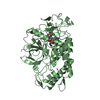
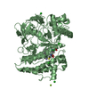
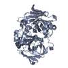
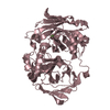
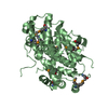
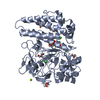
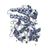
The biological assembly is a monomer. This was confirmed by size exclusion chromatography. The asymmetric unit contains two biological assemblies

-
Components

-
Protein , 1 types, 2 molecules AB

#1: Protein Sulfoxide synthase EgtB


Mass: 49562.574 Da / Num. of mol.: 2 / Fragment: UNP residues 3-446
Source method: isolated from a genetically manipulated source
Details: The N-terminal residues GH are left overs from an N-terminal TEV protease cleavage site. Some other N- and C-terminal residues were not visible in the electron density and have not been built.
Source: (gene. exp.) Mycobacterium thermoresistibile ATCC 19527 (bacteria)
Gene: KEK_08772 / Plasmid: pET19m / Production host: Escherichia coli BL21(DE3) (bacteria) / References: UniProt: G7CFI3

-
Non-polymers , 6 types, 1221 molecules

#2: Chemical
ChemComp-GOL / GLYCEROL / GLYCERIN / PROPANE-1,2,3-TRIOL


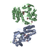
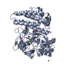
Mass: 92.094 Da / Num. of mol.: 10 / Source method: obtained synthetically / Formula: C3H8O3
#3: Chemical ChemComp-CL / CHLORIDE ION


Mass: 35.453 Da / Num. of mol.: 2 / Source method: obtained synthetically / Formula: Cl
#4: Chemical ChemComp-MG / MAGNESIUM ION


Mass: 24.305 Da / Num. of mol.: 2 / Source method: obtained synthetically / Formula: Mg
#5: Chemical ChemComp-FE / FE (III) ION


Mass: 55.845 Da / Num. of mol.: 2 / Source method: obtained synthetically / Formula: Fe
#6: Chemical ChemComp-CA / CALCIUM ION


Mass: 40.078 Da / Num. of mol.: 2 / Source method: obtained synthetically / Formula: Ca
#7: Water ChemComp-HOH / water


Mass: 18.015 Da / Num. of mol.: 1203 / Source method: isolated from a natural source / Formula: H2O

-
Experimental details

-
Experiment

ExperimentMethod: X-RAY DIFFRACTION / Number of used crystals: 1

-
Sample preparation

CrystalDensity Matthews: 3.23 Å3/Da / Density % sol: 61.97 %
Crystal growTemperature: 293 K / Method: vapor diffusion, hanging drop
Details: 0.1 M Tris/HCl pH 7-7.5, 6-12% PEG 8000, 0.2 M magnesium chloride

-
Data collection

Diffraction
IDMean temperature (K)Crystal-ID
11001
21001
Diffraction source
SourceSiteBeamlineIDWavelength (Å)
SYNCHROTRONSLS X06DA12.066
SYNCHROTRONSLS X06DA21
Detector
TypeIDDetectorDate
DECTRIS PILATUS 2M-F1PIXELFeb 14, 2014
DECTRIS PILATUS 2M-F2PIXELFeb 14, 2014
Radiation
IDProtocolMonochromatic (M) / Laue (L)Scattering typeWavelength-ID
1SINGLE WAVELENGTHMx-ray1
2SINGLE WAVELENGTHMx-ray2
Radiation wavelength
IDWavelength (Å)Relative weight
12.0661
211
ReflectionRedundancy: 43.6 % / Number: 2559724 / Rmerge(I) obs: 0.059 / D res high: 2.27 Å / D res low: 48.83 Å / Num. obs: 58661 / % possible obs: 96.4
Diffraction reflection shell
Highest resolution (Å)Lowest resolution (Å)IDRmerge(I) obsRedundancy
2.272.3310.29520.9
9.948.8310.02949.4
ReflectionResolution: 1.7→48.75 Å / Num. obs: 142371 / % possible obs: 100 % / Redundancy: 26.6 % / Biso Wilson estimate: 19.98 Å2 / CC1/2: 1 / Rmerge(I) obs: 0.118 / Rpim(I) all: 0.023 / Net I/σ(I): 24.6 / Num. measured all: 3781286 / Scaling rejects: 4
Reflection shell

Diffraction-ID: 1 / Rejects: _

Resolution (Å)Redundancy (%)Rmerge(I) obsMean I/σ(I) obsNum. measured allNum. unique allCC1/2Rpim(I) all% possible all
1.7-1.7327.51.5782.719159169660.7850.306100
9.31-48.7523.50.02887.124060102210.00699.4

-
Phasing

PhasingMethod: SAD

-
Processing

Software
NameVersionClassification
Aimless0.2.17data scaling
SHELXDEphasing
PHENIXrefinement
PDB_EXTRACT3.15data extraction
XDSdata reduction
ARPmodel building
Cootmodel building
RefinementMethod to determine structure: SAD / Resolution: 1.7→48.748 Å / SU ML: 0.13 / Cross valid method: FREE R-VALUE / σ(F): 1.36 / Phase error: 13.66 / Stereochemistry target values: ML
RfactorNum. reflection% reflectionSelection details
Rfree0.1569 7009 4.93 %random selection
Rwork0.1377 135254 --
obs0.1387 142263 99.99 %-
Solvent computationShrinkage radii: 0.9 Å / VDW probe radii: 1.11 Å / Solvent model: FLAT BULK SOLVENT MODEL
Displacement parametersBiso max: 92.93 Å2 / Biso mean: 28.9402 Å2 / Biso min: 10.4 Å2
Refinement stepCycle: final / Resolution: 1.7→48.748 Å
ProteinNucleic acidLigandSolventTotal
Num. atoms6708 0 162 1203 8073
Biso mean--41.01 40.13 -
Num. residues----854
Refine LS restraints
Refine-IDTypeDev idealNumber
X-RAY DIFFRACTIONf_bond_d0.0097064
X-RAY DIFFRACTIONf_angle_d1.2149659
X-RAY DIFFRACTIONf_chiral_restr0.046979
X-RAY DIFFRACTIONf_plane_restr0.0071293
X-RAY DIFFRACTIONf_dihedral_angle_d13.2882597
LS refinement shell

Refine-ID: X-RAY DIFFRACTION / Total num. of bins used: 30 / % reflection obs: 100 %

Resolution (Å)Rfactor RfreeNum. reflection RfreeRfactor RworkNum. reflection RworkNum. reflection all
1.7-1.71930.21792530.20144114664
1.7193-1.73960.22792650.197544154680
1.7396-1.76080.21042260.187244594685
1.7608-1.78310.19992340.184644524686
1.7831-1.80650.18352460.179644454691
1.8065-1.83130.2062100.169544884698
1.8313-1.85740.18782450.170844294674
1.8574-1.88520.19772270.164544794706
1.8852-1.91460.18362440.156744514695
1.9146-1.9460.16662560.147844674723
1.946-1.97960.14992220.144344534675
1.9796-2.01560.17072090.138545104719
2.0156-2.05430.16632500.135844514701
2.0543-2.09630.1682130.134144764689
2.0963-2.14180.15482400.133145064746
2.1418-2.19170.16672220.129444494671
2.1917-2.24650.14282400.129545104750
2.2465-2.30720.14871960.125844954691
2.3072-2.37510.16982210.126545254746
2.3751-2.45180.13642360.125544974733
2.4518-2.53940.15072170.122545344751
2.5394-2.64110.15022200.129445324752
2.6411-2.76120.13882350.128645084743
2.7612-2.90680.1452480.133744964744
2.9068-3.08890.16762350.130445434778
3.0889-3.32730.16432180.132545814799
3.3273-3.66210.14332510.132245674818
3.6621-4.19170.12861920.118646434835
4.1917-5.28020.12792500.121446574907
5.2802-48.76790.17752880.172848255113
Refinement TLS params.

Method: refined / Refine-ID: X-RAY DIFFRACTION

IDL112)L122)L132)L222)L232)L332)S11 (Å °)S12 (Å °)S13 (Å °)S21 (Å °)S22 (Å °)S23 (Å °)S31 (Å °)S32 (Å °)S33 (Å °)T112)T122)T132)T222)T232)T332)Origin x (Å)Origin y (Å)Origin z (Å)
11.37470.3338-0.02181.85590.30023.2618-0.03150.0234-0.1639-0.0874-0.02680.11370.1299-0.13480.04610.1087-0.010.02660.07740.02380.17823.070649.96742.2529
20.90690.3456-0.15391.2403-0.21510.72480.02390.01260.00350.0054-0.0469-0.0695-0.0380.10790.02450.0904-0.0040.01220.10780.02570.112237.688373.04137.8866
31.39540.34090.37871.8122-0.02861.17970.0607-0.09310.03880.0694-0.02630.2627-0.0034-0.1758-0.04370.0939-0.00160.02210.10560.02770.137525.995271.976839.2651
42.11780.1201-0.01351.77270.05032.28610.01330.06360.044-0.0690.0253-0.41030.030.499-0.04450.15190.00430.01240.2639-0.01330.227860.915227.8923.7514
51.1734-0.2107-0.22471.71790.4571.53030.03630.1954-0.0617-0.2682-0.00980.0641-0.028-0.1524-0.02050.1820.0195-0.010.19030.0010.127733.837722.778418.5229
62.6851-0.4373-0.91292.28740.46731.993-0.0368-0.0045-0.2398-0.11660.0638-0.11250.27210.1446-0.02770.21070.02340.00250.1564-0.02510.136542.665316.341719.4301
Refinement TLS group
IDRefine-IDRefine TLS-IDSelection detailsAuth asym-IDAuth seq-ID
1X-RAY DIFFRACTION1chain 'A' and (resid 7 through 125 )A0
2X-RAY DIFFRACTION2chain 'A' and (resid 126 through 365 )A0
3X-RAY DIFFRACTION3chain 'A' and (resid 366 through 434 )A0
4X-RAY DIFFRACTION4chain 'B' and (resid 8 through 148 )B0
5X-RAY DIFFRACTION5chain 'B' and (resid 149 through 345 )B0
6X-RAY DIFFRACTION6chain 'B' and (resid 346 through 433 )B0

+
About Yorodumi

-
News

-
Feb 9, 2022. New format data for meta-information of EMDB entries

New format data for meta-information of EMDB entries

  • Version 3 of the EMDB header file is now the official format.
  • The previous official version 1.9 will be removed from the archive.

Related info.:EMDB header

External links:wwPDB to switch to version 3 of the EMDB data model

-
Aug 12, 2020. Covid-19 info

Covid-19 info

URL: https://pdbjlvh1.pdbj.org/emnavi/covid19.php

New page: Covid-19 featured information page in EM Navigator.

Related info.:Covid-19 info / Mar 5, 2020. Novel coronavirus structure data

+
Mar 5, 2020. Novel coronavirus structure data

Novel coronavirus structure data

Related info.:Yorodumi Speices / Aug 12, 2020. Covid-19 info

External links:COVID-19 featured content - PDBj / Molecule of the Month (242):Coronavirus Proteases

+
Jan 31, 2019. EMDB accession codes are about to change! (news from PDBe EMDB page)

EMDB accession codes are about to change! (news from PDBe EMDB page)

  • The allocation of 4 digits for EMDB accession codes will soon come to an end. Whilst these codes will remain in use, new EMDB accession codes will include an additional digit and will expand incrementally as the available range of codes is exhausted. The current 4-digit format prefixed with “EMD-” (i.e. EMD-XXXX) will advance to a 5-digit format (i.e. EMD-XXXXX), and so on. It is currently estimated that the 4-digit codes will be depleted around Spring 2019, at which point the 5-digit format will come into force.
  • The EM Navigator/Yorodumi systems omit the EMD- prefix.

Related info.:Q: What is EMD? / ID/Accession-code notation in Yorodumi/EM Navigator

External links:EMDB Accession Codes are Changing Soon! / Contact to PDBj

+
Jul 12, 2017. Major update of PDB

Major update of PDB

  • wwPDB released updated PDB data conforming to the new PDBx/mmCIF dictionary.
  • This is a major update changing the version number from 4 to 5, and with Remediation, in which all the entries are updated.
  • In this update, many items about electron microscopy experimental information are reorganized (e.g. em_software).
  • Now, EM Navigator and Yorodumi are based on the updated data.

External links:wwPDB Remediation / Enriched Model Files Conforming to OneDep Data Standards Now Available in the PDB FTP Archive

-
Yorodumi

Thousand views of thousand structures

  • Yorodumi is a browser for structure data from EMDB, PDB, SASBDB, etc.
  • This page is also the successor to EM Navigator detail page, and also detail information page/front-end page for Omokage search.
  • The word "yorodu" (or yorozu) is an old Japanese word meaning "ten thousand". "mi" (miru) is to see.

Related info.:EMDB / PDB / SASBDB / Comparison of 3 databanks / Yorodumi Search / Aug 31, 2016. New EM Navigator & Yorodumi / Yorodumi Papers / Jmol/JSmol / Function and homology information / Changes in new EM Navigator and Yorodumi

Read more