 Movie
Movie Controller

[English] 日本語
 Yorodumi
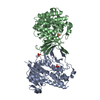
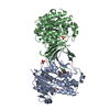
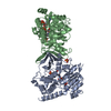
- PDB-4ooe: M. tuberculosis 1-deoxy-d-xylulose-5-phosphate reductoisomerase W... -
+ Open data

- Basic information
| Entry | Database: PDB / ID: 4ooe | ||||||
|---|---|---|---|---|---|---|---|
| Title | M. tuberculosis 1-deoxy-d-xylulose-5-phosphate reductoisomerase W203Y mutant bound to fosmidomycin and NADPH | ||||||
 Components Components | 1-deoxy-D-xylulose 5-phosphate reductoisomerase | ||||||
 Keywords Keywords | OXIDOREDUCTASE/ANTIBIOTIC / reductoisomerase / OXIDOREDUCTASE-ANTIBIOTIC complex | ||||||
| Function / homology |  Function and homology information Function and homology information: / : / 1-deoxy-D-xylulose-5-phosphate reductoisomerase / 1-deoxy-D-xylulose-5-phosphate reductoisomerase activity / cobalt ion binding / NADPH binding / manganese ion binding / magnesium ion binding Similarity search - Function | ||||||
| Biological species |   Mycobacterium tuberculosis (bacteria) Mycobacterium tuberculosis (bacteria) | ||||||
| Method |  X-RAY DIFFRACTION / SYNCHROTRON / MOLECULAR REPLACEMENT / Resolution: 1.826 Å X-RAY DIFFRACTION / SYNCHROTRON / MOLECULAR REPLACEMENT / Resolution: 1.826 Å | ||||||
 Authors Authors | Allen, C.L. / Kholodar, S.A. / Murkin, A.S. / Gulick, A.M. | ||||||
 Citation Citation | Journal: Biochemistry / Year: 2014 Title: Alteration of the Flexible Loop in 1-Deoxy-d-xylulose-5-phosphate Reductoisomerase Boosts Enthalpy-Driven Inhibition by Fosmidomycin. Authors: Kholodar, S.A. / Tombline, G. / Liu, J. / Tan, Z. / Allen, C.L. / Gulick, A.M. / Murkin, A.S. | ||||||
| History |
|
- Structure visualization
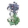
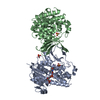
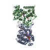
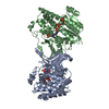
| Structure viewer | Molecule: MolmilJmol/JSmol |
|---|
- Downloads & links
-Download
| PDBx/mmCIF format | 4ooe.cif.gz | 330.6 KB | Display | PDBx/mmCIF format |
|---|---|---|---|---|
| PDB format | pdb4ooe.ent.gz | 264.8 KB | Display | PDB format |
| PDBx/mmJSON format | 4ooe.json.gz | Tree view | PDBx/mmJSON format | |
| Others |  Other downloads Other downloads |
-Validation report
| Arichive directory | https://data.pdbj.org/pub/pdb/validation_reports/oo/4ooeftp://data.pdbj.org/pub/pdb/validation_reports/oo/4ooe | HTTPS FTP |
|---|
-Related structure data
| Related structure data |  4oofC 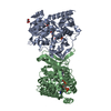 4a03S C: citing same article ( S: Starting model for refinement |
|---|---|
| Similar structure data |


-Links
 PDBj
PDBj

- Assembly
| Deposited unit | 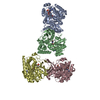
| ||||||||
|---|---|---|---|---|---|---|---|---|---|
| 1 | 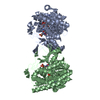
| ||||||||
| 2 | 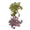
| ||||||||
| Unit cell |
|
-Components
| #1: Protein | Mass: 42443.879 Da / Num. of mol.: 4 / Fragment: UNP residues 1-389 / Mutation: W203Y Source method: isolated from a genetically manipulated source Source: (gene. exp.) Mycobacterium tuberculosis (bacteria) / Strain: H37Rv / Gene: dxr, RVBD_2870c / Production host: References: UniProt: I6YAH0, UniProt: P9WNS1*PLUS, 1-deoxy-D-xylulose-5-phosphate reductoisomerase #2: Chemical | ChemComp-FOM / 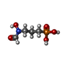  Mass: 183.100 Da / Num. of mol.: 4 / Source method: obtained synthetically / Formula: C4H10NO5P / Comment: antibiotic*YM Mass: 183.100 Da / Num. of mol.: 4 / Source method: obtained synthetically / Formula: C4H10NO5P / Comment: antibiotic*YM#3: Chemical | ChemComp-MN /   Mass: 54.938 Da / Num. of mol.: 4 / Source method: obtained synthetically / Formula: Mn Mass: 54.938 Da / Num. of mol.: 4 / Source method: obtained synthetically / Formula: Mn#4: Chemical | ChemComp-NDP /   Mass: 745.421 Da / Num. of mol.: 4 / Source method: obtained synthetically / Formula: C21H30N7O17P3 Mass: 745.421 Da / Num. of mol.: 4 / Source method: obtained synthetically / Formula: C21H30N7O17P3#5: Water | ChemComp-HOH / |  Mass: 18.015 Da / Num. of mol.: 1671 / Source method: isolated from a natural source / Formula: H2O Mass: 18.015 Da / Num. of mol.: 1671 / Source method: isolated from a natural source / Formula: H2O |
|---|
-Experimental details
-Experiment
| Experiment | Method: X-RAY DIFFRACTION / Number of used crystals: 1 |
|---|
- Sample preparation
| Crystal | Density Matthews: 2.52 Å3/Da / Density % sol: 51.1 % |
|---|---|
| Crystal grow | Temperature: 293 K / Method: vapor diffusion, hanging drop / pH: 5.5 Details: 6% PEG4000, 50 mM MES, 20% MPD, pH 5.5, VAPOR DIFFUSION, HANGING DROP, temperature 293K |
-Data collection
| Diffraction | Mean temperature: 113 K |
|---|---|
| Diffraction source | Source: SYNCHROTRON / Site: SSRL  / Beamline: BL9-2 / Wavelength: 1.284 Å / Beamline: BL9-2 / Wavelength: 1.284 Å |
| Detector | Type: MARMOSAIC 325 mm CCD / Detector: CCD / Date: Jan 11, 2013 |
| Radiation | Monochromator: Double crystal Si(111) / Protocol: SINGLE WAVELENGTH / Monochromatic (M) / Laue (L): M / Scattering type: x-ray |
| Radiation wavelength | Wavelength: 1.284 Å / Relative weight: 1 |
| Reflection | Resolution: 1.826→86.712 Å / Num. all: 151359 / Num. obs: 145456 / % possible obs: 96.1 % / Observed criterion σ(F): 0 / Observed criterion σ(I): 0 / Redundancy: 3.1 % / Biso Wilson estimate: 18.5 Å2 / Rmerge(I) obs: 0.061 / Net I/σ(I): 16 |
| Reflection shell | Resolution: 1.826→1.93 Å / Redundancy: 2.7 % / Rmerge(I) obs: 0.247 / Mean I/σ(I) obs: 3.8 / Num. unique all: 18847 / % possible all: 86.1 |
- Processing
| Software |
| |||||||||||||||||||||||||||||||||||||||||||||||||||||||||||||||||||||||||||||||||||||||||||||||||||||||||
|---|---|---|---|---|---|---|---|---|---|---|---|---|---|---|---|---|---|---|---|---|---|---|---|---|---|---|---|---|---|---|---|---|---|---|---|---|---|---|---|---|---|---|---|---|---|---|---|---|---|---|---|---|---|---|---|---|---|---|---|---|---|---|---|---|---|---|---|---|---|---|---|---|---|---|---|---|---|---|---|---|---|---|---|---|---|---|---|---|---|---|---|---|---|---|---|---|---|---|---|---|---|---|---|---|---|---|
| Refinement | Method to determine structure: MOLECULAR REPLACEMENT Starting model: PDB ENTRY 4A03 Resolution: 1.826→38.827 Å / SU ML: 0.19 / σ(F): 0 / Phase error: 20.63 / Stereochemistry target values: ML
| |||||||||||||||||||||||||||||||||||||||||||||||||||||||||||||||||||||||||||||||||||||||||||||||||||||||||
| Solvent computation | Shrinkage radii: 0.9 Å / VDW probe radii: 1.11 Å / Solvent model: FLAT BULK SOLVENT MODEL | |||||||||||||||||||||||||||||||||||||||||||||||||||||||||||||||||||||||||||||||||||||||||||||||||||||||||
| Refinement step | Cycle: LAST / Resolution: 1.826→38.827 Å
| |||||||||||||||||||||||||||||||||||||||||||||||||||||||||||||||||||||||||||||||||||||||||||||||||||||||||
| Refine LS restraints |
| |||||||||||||||||||||||||||||||||||||||||||||||||||||||||||||||||||||||||||||||||||||||||||||||||||||||||
| LS refinement shell |
|
