 Movie
Movie Controller

[English] 日本語
 Yorodumi
- PDB-4a03: Crystal Structure of Mycobacterium tuberculosis DXR in complex wi... -
+ Open data

- Basic information
| Entry | Database: PDB / ID: 4a03 | ||||||
|---|---|---|---|---|---|---|---|
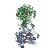
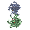
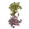
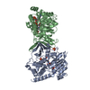
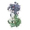
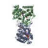
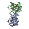
| Title | Crystal Structure of Mycobacterium tuberculosis DXR in complex with the antibiotic FR900098 and cofactor NADPH | ||||||
 Components Components | 1-DEOXY-D-XYLULOSE 5-PHOSPHATE REDUCTOISOMERASE | ||||||
 Keywords Keywords | OXIDOREDUCTASE / FR900098 / MEP PATHWAY | ||||||
| Function / homology |  Function and homology information Function and homology information: / : / 1-deoxy-D-xylulose-5-phosphate reductoisomerase / 1-deoxy-D-xylulose-5-phosphate reductoisomerase activity / cobalt ion binding / NADPH binding / manganese ion binding / magnesium ion binding Similarity search - Function | ||||||
| Biological species |   MYCOBACTERIUM TUBERCULOSIS (bacteria) MYCOBACTERIUM TUBERCULOSIS (bacteria) | ||||||
| Method |  X-RAY DIFFRACTION / SYNCHROTRON / MOLECULAR REPLACEMENT / Resolution: 1.65 Å X-RAY DIFFRACTION / SYNCHROTRON / MOLECULAR REPLACEMENT / Resolution: 1.65 Å | ||||||
 Authors Authors | Bjorkelid, C. / Bergfors, T. / Jones, T.A. | ||||||
 Citation Citation | Journal: Acta Crystallogr.,Sect.D / Year: 2012 Title: Structural Studies on Mycobacterium Tuberculosis Dxr in Complex with the Antibiotic Fr-900098. Authors: Bjorkelid, C. / Bergfors, T. / Unge, T. / Mowbray, S.L. / Jones, T.A. | ||||||
| History |
|
- Structure visualization
| Structure viewer | Molecule: MolmilJmol/JSmol |
|---|
- Downloads & links
-Download
| PDBx/mmCIF format | 4a03.cif.gz | 167.6 KB | Display | PDBx/mmCIF format |
|---|---|---|---|---|
| PDB format | pdb4a03.ent.gz | 130.9 KB | Display | PDB format |
| PDBx/mmJSON format | 4a03.json.gz | Tree view | PDBx/mmJSON format | |
| Others |  Other downloads Other downloads |
-Validation report
| Arichive directory | https://data.pdbj.org/pub/pdb/validation_reports/a0/4a03ftp://data.pdbj.org/pub/pdb/validation_reports/a0/4a03 | HTTPS FTP |
|---|
-Related structure data
| Related structure data |  2jcz S: Starting model for refinement |
|---|---|
| Similar structure data |


-Links
 PDBj
PDBj

- Assembly
| Deposited unit | 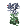
| ||||||||
|---|---|---|---|---|---|---|---|---|---|
| 1 |
| ||||||||
| Unit cell |
| ||||||||
| Noncrystallographic symmetry (NCS) | NCS oper: (Code: given Matrix: (0.15, -0.0909, -0.9845), Vector: |
-Components
-Protein , 1 types, 2 molecules AB
| #1: Protein | Mass: 41700.973 Da / Num. of mol.: 2 / Fragment: RESIDUES 2-389 Source method: isolated from a genetically manipulated source Source: (gene. exp.) MYCOBACTERIUM TUBERCULOSIS (bacteria) / Strain: H37RV / Plasmid: PEXP5CT/TOPO / Production host: References: UniProt: P64012, UniProt: P9WNS1*PLUS, 1-deoxy-D-xylulose-5-phosphate reductoisomerase |
|---|
-Non-polymers , 5 types, 536 molecules 


| #2: Chemical |  Mass: 54.938 Da / Num. of mol.: 2 / Source method: obtained synthetically / Formula: Mn Mass: 54.938 Da / Num. of mol.: 2 / Source method: obtained synthetically / Formula: Mn#3: Chemical |  Mass: 197.126 Da / Num. of mol.: 2 / Source method: obtained synthetically / Formula: C5H12NO5P Mass: 197.126 Da / Num. of mol.: 2 / Source method: obtained synthetically / Formula: C5H12NO5P#4: Chemical |  Mass: 745.421 Da / Num. of mol.: 2 / Source method: obtained synthetically / Formula: C21H30N7O17P3 Mass: 745.421 Da / Num. of mol.: 2 / Source method: obtained synthetically / Formula: C21H30N7O17P3#5: Chemical | ChemComp-GOL / |  Mass: 92.094 Da / Num. of mol.: 1 / Source method: obtained synthetically / Formula: C3H8O3 Mass: 92.094 Da / Num. of mol.: 1 / Source method: obtained synthetically / Formula: C3H8O3#6: Water | ChemComp-HOH / | Mass: 18.015 Da / Num. of mol.: 529 / Source method: isolated from a natural source / Formula: H2O |
|---|
-Experimental details
-Experiment
| Experiment | Method: X-RAY DIFFRACTION / Number of used crystals: 1 |
|---|
- Sample preparation
| Crystal | Density Matthews: 2.44 Å3/Da / Density % sol: 49.66 % / Description: NONE |
|---|---|
| Crystal grow | Temperature: 293 K / Method: vapor diffusion, sitting drop Details: SITTING-DROP VAPOR-DIFFUSION METHOD AT 293 K. 10% (W/V) PEG 20000, 20% (V/V) MONOMETHYL ETHER PEG 550, 0.1 M MES/IMIDAZOLE, PH 6.5, AND 0.03 M EACH OF DIETHYLENEGLYCOL, TRIETHYLENEGLYCOL, ...Details: SITTING-DROP VAPOR-DIFFUSION METHOD AT 293 K. 10% (W/V) PEG 20000, 20% (V/V) MONOMETHYL ETHER PEG 550, 0.1 M MES/IMIDAZOLE, PH 6.5, AND 0.03 M EACH OF DIETHYLENEGLYCOL, TRIETHYLENEGLYCOL, TETRAETHYLENEGLYCOL AND PENTAETHYLENEGLYCOL. |
-Data collection
| Diffraction | Mean temperature: 110 K |
|---|---|
| Diffraction source | Source: SYNCHROTRON / Site: ESRF  / Beamline: ID23-1 / Wavelength: 0.9768 / Beamline: ID23-1 / Wavelength: 0.9768 |
| Detector | Type: ADSC QUANTUM 315r / Detector: CCD / Date: Sep 17, 2010 |
| Radiation | Protocol: SINGLE WAVELENGTH / Monochromatic (M) / Laue (L): M / Scattering type: x-ray |
| Radiation wavelength | Wavelength: 0.9768 Å / Relative weight: 1 |
| Reflection | Resolution: 1.65→42.21 Å / Num. obs: 101922 / % possible obs: 99.8 % / Observed criterion σ(I): 0 / Redundancy: 7.8 % / Rmerge(I) obs: 0.09 / Net I/σ(I): 14.4 |
| Reflection shell | Resolution: 1.65→1.74 Å / Redundancy: 7.9 % / Rmerge(I) obs: 0.46 / Mean I/σ(I) obs: 4.6 / % possible all: 100 |
- Processing
| Software |
| ||||||||||||||||||||||||||||||||||||||||||||||||||||||||||||||||||||||||||||||||||||||||||||||||||||||||||||||||||||||||||||||||||||||||||||||||||||||||||||||||||||||||||||||||||||||
|---|---|---|---|---|---|---|---|---|---|---|---|---|---|---|---|---|---|---|---|---|---|---|---|---|---|---|---|---|---|---|---|---|---|---|---|---|---|---|---|---|---|---|---|---|---|---|---|---|---|---|---|---|---|---|---|---|---|---|---|---|---|---|---|---|---|---|---|---|---|---|---|---|---|---|---|---|---|---|---|---|---|---|---|---|---|---|---|---|---|---|---|---|---|---|---|---|---|---|---|---|---|---|---|---|---|---|---|---|---|---|---|---|---|---|---|---|---|---|---|---|---|---|---|---|---|---|---|---|---|---|---|---|---|---|---|---|---|---|---|---|---|---|---|---|---|---|---|---|---|---|---|---|---|---|---|---|---|---|---|---|---|---|---|---|---|---|---|---|---|---|---|---|---|---|---|---|---|---|---|---|---|---|---|
| Refinement | Method to determine structure: MOLECULAR REPLACEMENT Starting model: PDB ENTRY 2JCZ 2jcz Resolution: 1.65→42.21 Å / Cor.coef. Fo:Fc: 0.956 / Cor.coef. Fo:Fc free: 0.943 / SU B: 1.659 / SU ML: 0.058 / Cross valid method: THROUGHOUT / ESU R: 0.094 / ESU R Free: 0.092 / Stereochemistry target values: MAXIMUM LIKELIHOOD / Details: HYDROGENS HAVE BEEN ADDED IN THE RIDING POSITIONS.
| ||||||||||||||||||||||||||||||||||||||||||||||||||||||||||||||||||||||||||||||||||||||||||||||||||||||||||||||||||||||||||||||||||||||||||||||||||||||||||||||||||||||||||||||||||||||
| Solvent computation | Ion probe radii: 0.8 Å / Shrinkage radii: 0.8 Å / VDW probe radii: 1.2 Å / Solvent model: MASK | ||||||||||||||||||||||||||||||||||||||||||||||||||||||||||||||||||||||||||||||||||||||||||||||||||||||||||||||||||||||||||||||||||||||||||||||||||||||||||||||||||||||||||||||||||||||
| Displacement parameters | Biso mean: 22.132 Å2
| ||||||||||||||||||||||||||||||||||||||||||||||||||||||||||||||||||||||||||||||||||||||||||||||||||||||||||||||||||||||||||||||||||||||||||||||||||||||||||||||||||||||||||||||||||||||
| Refinement step | Cycle: LAST / Resolution: 1.65→42.21 Å
| ||||||||||||||||||||||||||||||||||||||||||||||||||||||||||||||||||||||||||||||||||||||||||||||||||||||||||||||||||||||||||||||||||||||||||||||||||||||||||||||||||||||||||||||||||||||
| Refine LS restraints |
|