 Movie
Movie Controller

[English] 日本語
 Yorodumi
- PDB-3p96: Crystal structure of Phosphoserine phosphatase SerB from Mycobact... -
+ Open data

- Basic information
| Entry | Database: PDB / ID: 3p96 | ||||||
|---|---|---|---|---|---|---|---|
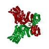
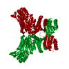
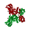
| Title | Crystal structure of Phosphoserine phosphatase SerB from Mycobacterium avium, native form | ||||||
 Components Components | Phosphoserine phosphatase SerB | ||||||
 Keywords Keywords | HYDROLASE / SSGCID / Phosphoserine phosphatase SerB / Structural Genomics / Seattle Structural Genomics Center for Infectious Disease | ||||||
| Function / homology |  Function and homology information Function and homology informationphosphoserine phosphatase / L-phosphoserine phosphatase activity / L-serine biosynthetic process / magnesium ion binding / cytoplasm Similarity search - Function | ||||||
| Biological species |  Mycobacterium avium (bacteria) Mycobacterium avium (bacteria) | ||||||
| Method |  X-RAY DIFFRACTION / SYNCHROTRON / SAD / Resolution: 2.05 Å X-RAY DIFFRACTION / SYNCHROTRON / SAD / Resolution: 2.05 Å | ||||||
 Authors Authors | Seattle Structural Genomics Center for Infectious Disease (SSGCID) | ||||||
 Citation Citation | Journal: J.Struct.Funct.Genom. / Year: 2011 Title: SAD phasing using iodide ions in a high-throughput structural genomics environment. Authors: Abendroth, J. / Gardberg, A.S. / Robinson, J.I. / Christensen, J.S. / Staker, B.L. / Myler, P.J. / Stewart, L.J. / Edwards, T.E. #1: Journal: Tuberculosis (Edinb) / Year: 2014Title: Increasing the structural coverage of tuberculosis drug targets. Authors: Baugh, L. / Phan, I. / Begley, D.W. / Clifton, M.C. / Armour, B. / Dranow, D.M. / Taylor, B.M. / Muruthi, M.M. / Abendroth, J. / Fairman, J.W. / Fox, D. / Dieterich, S.H. / Staker, B.L. / ...Authors: Baugh, L. / Phan, I. / Begley, D.W. / Clifton, M.C. / Armour, B. / Dranow, D.M. / Taylor, B.M. / Muruthi, M.M. / Abendroth, J. / Fairman, J.W. / Fox, D. / Dieterich, S.H. / Staker, B.L. / Gardberg, A.S. / Choi, R. / Hewitt, S.N. / Napuli, A.J. / Myers, J. / Barrett, L.K. / Zhang, Y. / Ferrell, M. / Mundt, E. / Thompkins, K. / Tran, N. / Lyons-Abbott, S. / Abramov, A. / Sekar, A. / Serbzhinskiy, D. / Lorimer, D. / Buchko, G.W. / Stacy, R. / Stewart, L.J. / Edwards, T.E. / Van Voorhis, W.C. / Myler, P.J. | ||||||
| History |
|
- Structure visualization
| Structure viewer | Molecule: MolmilJmol/JSmol |
|---|
- Downloads & links
-Download
| PDBx/mmCIF format | 3p96.cif.gz | 162 KB | Display | PDBx/mmCIF format |
|---|---|---|---|---|
| PDB format | pdb3p96.ent.gz | 129.6 KB | Display | PDB format |
| PDBx/mmJSON format | 3p96.json.gz | Tree view | PDBx/mmJSON format | |
| Others |  Other downloads Other downloads |
-Validation report
| Arichive directory | https://data.pdbj.org/pub/pdb/validation_reports/p9/3p96ftp://data.pdbj.org/pub/pdb/validation_reports/p9/3p96 | HTTPS FTP |
|---|
-Related structure data
| Related structure data |  3k9gC  3km3C  3kw3C  3luzC  3menC  3njbC  3o2eC 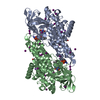 3oibC  3pfdC 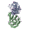 3pm6C C: citing same article ( |
|---|---|
| Similar structure data | |
| Other databases |
-Links
 PDBj
PDBj

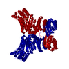
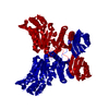
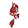
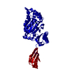
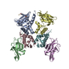
- Assembly
| Deposited unit | 
| ||||||||
|---|---|---|---|---|---|---|---|---|---|
| 1 | 
| ||||||||
| Unit cell |
|
-Components
| #1: Protein | Mass: 43852.094 Da / Num. of mol.: 1 Source method: isolated from a genetically manipulated source Source: (gene. exp.) Mycobacterium avium (bacteria) / Strain: 104 / Gene: serB, MAV_3907 / Plasmid: AVA0421 / Production host: |
|---|---|
| #2: Chemical | ChemComp-MG /   Mass: 24.305 Da / Num. of mol.: 1 / Source method: obtained synthetically / Formula: Mg Mass: 24.305 Da / Num. of mol.: 1 / Source method: obtained synthetically / Formula: Mg |
| #3: Chemical | ChemComp-CL /   Mass: 35.453 Da / Num. of mol.: 1 / Source method: obtained synthetically / Formula: Cl Mass: 35.453 Da / Num. of mol.: 1 / Source method: obtained synthetically / Formula: Cl |
| #4: Chemical | ChemComp-EDO /   Mass: 62.068 Da / Num. of mol.: 1 / Source method: obtained synthetically / Formula: C2H6O2 Mass: 62.068 Da / Num. of mol.: 1 / Source method: obtained synthetically / Formula: C2H6O2 |
| #5: Water | ChemComp-HOH /  Mass: 18.015 Da / Num. of mol.: 112 / Source method: isolated from a natural source / Formula: H2O Mass: 18.015 Da / Num. of mol.: 112 / Source method: isolated from a natural source / Formula: H2O |
| Sequence details | AUTHORS STATE THAT THE UNIPROT SEQUENCE #A0QJI1 IS BEING UPDATED |
-Experimental details
-Experiment
| Experiment | Method: X-RAY DIFFRACTION / Number of used crystals: 1 |
|---|
- Sample preparation
| Crystal | Density Matthews: 2.6 Å3/Da / Density % sol: 52.69 % |
|---|---|
| Crystal grow | Temperature: 290 K / Method: vapor diffusion, sitting drop / pH: 6 Details: 0.2 M MgCl2, 0.1 M MES, 20% PEG6000, protein at 27.45mg/ml, pH 6.0, VAPOR DIFFUSION, SITTING DROP, temperature 290K |
-Data collection
| Diffraction | Mean temperature: 100 K | ||||||||||||||||||||||||||||||||||||||||||||||||||||||||||||||||||||||||||||||||||||||||||||||||||||||||||||||||||||||||||||||||||||||||||||||||||||||||||||||||||||||||
|---|---|---|---|---|---|---|---|---|---|---|---|---|---|---|---|---|---|---|---|---|---|---|---|---|---|---|---|---|---|---|---|---|---|---|---|---|---|---|---|---|---|---|---|---|---|---|---|---|---|---|---|---|---|---|---|---|---|---|---|---|---|---|---|---|---|---|---|---|---|---|---|---|---|---|---|---|---|---|---|---|---|---|---|---|---|---|---|---|---|---|---|---|---|---|---|---|---|---|---|---|---|---|---|---|---|---|---|---|---|---|---|---|---|---|---|---|---|---|---|---|---|---|---|---|---|---|---|---|---|---|---|---|---|---|---|---|---|---|---|---|---|---|---|---|---|---|---|---|---|---|---|---|---|---|---|---|---|---|---|---|---|---|---|---|---|---|---|---|---|
| Diffraction source | Source: SYNCHROTRON / Site: ALS  / Beamline: 5.0.1 / Wavelength: 0.9774 Å / Beamline: 5.0.1 / Wavelength: 0.9774 Å | ||||||||||||||||||||||||||||||||||||||||||||||||||||||||||||||||||||||||||||||||||||||||||||||||||||||||||||||||||||||||||||||||||||||||||||||||||||||||||||||||||||||||
| Detector | Type: ADSC QUANTUM 315 / Detector: CCD / Date: Sep 15, 2010 | ||||||||||||||||||||||||||||||||||||||||||||||||||||||||||||||||||||||||||||||||||||||||||||||||||||||||||||||||||||||||||||||||||||||||||||||||||||||||||||||||||||||||
| Radiation | Protocol: SINGLE WAVELENGTH / Monochromatic (M) / Laue (L): M / Scattering type: x-ray | ||||||||||||||||||||||||||||||||||||||||||||||||||||||||||||||||||||||||||||||||||||||||||||||||||||||||||||||||||||||||||||||||||||||||||||||||||||||||||||||||||||||||
| Radiation wavelength | Wavelength: 0.9774 Å / Relative weight: 1 | ||||||||||||||||||||||||||||||||||||||||||||||||||||||||||||||||||||||||||||||||||||||||||||||||||||||||||||||||||||||||||||||||||||||||||||||||||||||||||||||||||||||||
| Reflection | Resolution: 2.05→36.97 Å / Num. all: 30644 / Num. obs: 29584 / % possible obs: 96.5 % / Observed criterion σ(F): 0 / Observed criterion σ(I): -3 / Redundancy: 5.7 % / Biso Wilson estimate: 38.011 Å2 / Rmerge(I) obs: 0.062 / Net I/σ(I): 16.41 | ||||||||||||||||||||||||||||||||||||||||||||||||||||||||||||||||||||||||||||||||||||||||||||||||||||||||||||||||||||||||||||||||||||||||||||||||||||||||||||||||||||||||
| Reflection shell | Diffraction-ID: 1
|
- Processing
| Software |
| ||||||||||||||||||||||||||||||||||||||||||||||||||||||||||||||||||||||||||||||||||||||||||||||||||||||||||||||||||||||||||||||||||||||||||||||||||||||||||||||||||||||||||||||||||||||||||||||||||||||||
|---|---|---|---|---|---|---|---|---|---|---|---|---|---|---|---|---|---|---|---|---|---|---|---|---|---|---|---|---|---|---|---|---|---|---|---|---|---|---|---|---|---|---|---|---|---|---|---|---|---|---|---|---|---|---|---|---|---|---|---|---|---|---|---|---|---|---|---|---|---|---|---|---|---|---|---|---|---|---|---|---|---|---|---|---|---|---|---|---|---|---|---|---|---|---|---|---|---|---|---|---|---|---|---|---|---|---|---|---|---|---|---|---|---|---|---|---|---|---|---|---|---|---|---|---|---|---|---|---|---|---|---|---|---|---|---|---|---|---|---|---|---|---|---|---|---|---|---|---|---|---|---|---|---|---|---|---|---|---|---|---|---|---|---|---|---|---|---|---|---|---|---|---|---|---|---|---|---|---|---|---|---|---|---|---|---|---|---|---|---|---|---|---|---|---|---|---|---|---|---|---|---|
| Refinement | Method to determine structure: SAD / Resolution: 2.05→36.97 Å / Cor.coef. Fo:Fc: 0.945 / Cor.coef. Fo:Fc free: 0.91 / SU B: 16.038 / SU ML: 0.181 / Cross valid method: THROUGHOUT / σ(F): 0 / ESU R Free: 0.195 / Stereochemistry target values: MAXIMUM LIKELIHOOD / Details: HYDROGENS HAVE BEEN ADDED IN THE RIDING POSITIONS
| ||||||||||||||||||||||||||||||||||||||||||||||||||||||||||||||||||||||||||||||||||||||||||||||||||||||||||||||||||||||||||||||||||||||||||||||||||||||||||||||||||||||||||||||||||||||||||||||||||||||||
| Solvent computation | Ion probe radii: 0.8 Å / Shrinkage radii: 0.8 Å / VDW probe radii: 1.4 Å / Solvent model: MASK | ||||||||||||||||||||||||||||||||||||||||||||||||||||||||||||||||||||||||||||||||||||||||||||||||||||||||||||||||||||||||||||||||||||||||||||||||||||||||||||||||||||||||||||||||||||||||||||||||||||||||
| Displacement parameters | Biso mean: 45.015 Å2
| ||||||||||||||||||||||||||||||||||||||||||||||||||||||||||||||||||||||||||||||||||||||||||||||||||||||||||||||||||||||||||||||||||||||||||||||||||||||||||||||||||||||||||||||||||||||||||||||||||||||||
| Refinement step | Cycle: LAST / Resolution: 2.05→36.97 Å
| ||||||||||||||||||||||||||||||||||||||||||||||||||||||||||||||||||||||||||||||||||||||||||||||||||||||||||||||||||||||||||||||||||||||||||||||||||||||||||||||||||||||||||||||||||||||||||||||||||||||||
| Refine LS restraints |
| ||||||||||||||||||||||||||||||||||||||||||||||||||||||||||||||||||||||||||||||||||||||||||||||||||||||||||||||||||||||||||||||||||||||||||||||||||||||||||||||||||||||||||||||||||||||||||||||||||||||||
| LS refinement shell | Resolution: 2.05→2.103 Å / Total num. of bins used: 20
| ||||||||||||||||||||||||||||||||||||||||||||||||||||||||||||||||||||||||||||||||||||||||||||||||||||||||||||||||||||||||||||||||||||||||||||||||||||||||||||||||||||||||||||||||||||||||||||||||||||||||
| Refinement TLS params. | Method: refined / Refine-ID: X-RAY DIFFRACTION
| ||||||||||||||||||||||||||||||||||||||||||||||||||||||||||||||||||||||||||||||||||||||||||||||||||||||||||||||||||||||||||||||||||||||||||||||||||||||||||||||||||||||||||||||||||||||||||||||||||||||||
| Refinement TLS group |
|
