 Movie
Movie Controller

[English] 日本語
 Yorodumi
- PDB-3km3: Crystal structure of eoxycytidine triphosphate deaminase from ana... -
+ Open data

- Basic information
| Entry | Database: PDB / ID: 3km3 | ||||||
|---|---|---|---|---|---|---|---|
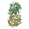
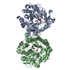
| Title | Crystal structure of eoxycytidine triphosphate deaminase from anaplasma phagocytophilum at 2.1A resolution | ||||||
 Components Components | Deoxycytidine triphosphate deaminase | ||||||
 Keywords Keywords | HYDROLASE / SSGCID / NIH / NIAID / SBRI / UW / DECODE / DEOXYCYTIDINE TRIPHOSPHATE DEAMINASE / ANAPLASMA PHAGOCYTOPHILUM / IODIDE PHASING / Nucleotide metabolism / Structural Genomics / Seattle Structural Genomics Center for Infectious Disease | ||||||
| Function / homology |  Function and homology information Function and homology informationdCTP deaminase / dUTP biosynthetic process / dCTP deaminase activity / dUMP biosynthetic process / nucleotide binding Similarity search - Function | ||||||
| Biological species |  Anaplasma phagocytophilum (agent of human granulocytic ehrlichiosis) Anaplasma phagocytophilum (agent of human granulocytic ehrlichiosis) | ||||||
| Method |  X-RAY DIFFRACTION / SAD / Resolution: 2.1 Å X-RAY DIFFRACTION / SAD / Resolution: 2.1 Å | ||||||
 Authors Authors | Seattle Structural Genomics Center for Infectious Disease (SSGCID) | ||||||
 Citation Citation | Journal: J.Struct.Funct.Genom. / Year: 2011 Title: SAD phasing using iodide ions in a high-throughput structural genomics environment. Authors: Abendroth, J. / Gardberg, A.S. / Robinson, J.I. / Christensen, J.S. / Staker, B.L. / Myler, P.J. / Stewart, L.J. / Edwards, T.E. | ||||||
| History |
|
- Structure visualization
| Structure viewer | Molecule: MolmilJmol/JSmol |
|---|
- Downloads & links
-Download
| PDBx/mmCIF format | 3km3.cif.gz | 81 KB | Display | PDBx/mmCIF format |
|---|---|---|---|---|
| PDB format | pdb3km3.ent.gz | 61 KB | Display | PDB format |
| PDBx/mmJSON format | 3km3.json.gz | Tree view | PDBx/mmJSON format | |
| Others |  Other downloads Other downloads |
-Validation report
| Arichive directory | https://data.pdbj.org/pub/pdb/validation_reports/km/3km3ftp://data.pdbj.org/pub/pdb/validation_reports/km/3km3 | HTTPS FTP |
|---|
-Related structure data
| Related structure data |  3k9gC  3kw3C  3luzC  3menC  3njbC  3o2eC 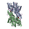 3oibC  3p96C  3pfdC 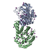 3pm6C C: citing same article ( |
|---|---|
| Similar structure data | |
| Other databases |


-Links
 PDBj
PDBj
- Assembly
| Deposited unit | 
| |||||||||||||||||||||||||||
|---|---|---|---|---|---|---|---|---|---|---|---|---|---|---|---|---|---|---|---|---|---|---|---|---|---|---|---|---|
| 1 | 
| |||||||||||||||||||||||||||
| Unit cell |
| |||||||||||||||||||||||||||
| Components on special symmetry positions |
| |||||||||||||||||||||||||||
| Noncrystallographic symmetry (NCS) | NCS domain:
NCS domain segments:
|
-Components
| #1: Protein | Mass: 22901.010 Da / Num. of mol.: 2 Source method: isolated from a genetically manipulated source Source: (gene. exp.) Anaplasma phagocytophilum (agent of human granulocytic ehrlichiosis)Strain: HZ / Gene: dcd, APH_0130 / Plasmid: AVA0421 / Production host: #2: Chemical | ChemComp-IOD /   Mass: 126.904 Da / Num. of mol.: 16 / Source method: obtained synthetically / Formula: I Mass: 126.904 Da / Num. of mol.: 16 / Source method: obtained synthetically / Formula: I#3: Water | ChemComp-HOH / |  Mass: 18.015 Da / Num. of mol.: 176 / Source method: isolated from a natural source / Formula: H2O Mass: 18.015 Da / Num. of mol.: 176 / Source method: isolated from a natural source / Formula: H2O |
|---|
-Experimental details
-Experiment
| Experiment | Method: X-RAY DIFFRACTION / Number of used crystals: 1 |
|---|
- Sample preparation
| Crystal | Density Matthews: 2.12 Å3/Da / Density % sol: 41.96 % |
|---|---|
| Crystal grow | Temperature: 290 K / pH: 8.1 Details: MD PACT SCREEN CONDITION D9: 100MM TRIS PH 8.1, 200MM LICL, 20% PEG 3350; ANPHA.00973.A AT XXMG/ ML, CRYSTAL SOAKED IN 100MM HEPES PH 7.0, 200MM NACL, 20% PEG 3350, 1M KI, VAPOR DIFFUSION, ...Details: MD PACT SCREEN CONDITION D9: 100MM TRIS PH 8.1, 200MM LICL, 20% PEG 3350; ANPHA.00973.A AT XXMG/ ML, CRYSTAL SOAKED IN 100MM HEPES PH 7.0, 200MM NACL, 20% PEG 3350, 1M KI, VAPOR DIFFUSION, SITTING DROP, TEMPERATURE 290K |
-Data collection
| Diffraction | Mean temperature: 100 K |
|---|---|
| Diffraction source | Source: ROTATING ANODE / Type: RIGAKU MICROMAX-007 HF / Wavelength: 1.5418 |
| Detector | Type: RIGAKU SATURN 944 / Detector: CCD / Date: Nov 5, 2009 |
| Radiation | Protocol: SINGLE WAVELENGTH / Monochromatic (M) / Laue (L): M / Scattering type: x-ray |
| Radiation wavelength | Wavelength: 1.5418 Å / Relative weight: 1 |
| Reflection | Resolution: 2.1→20 Å / Num. obs: 21725 / % possible obs: 98.9 % / Observed criterion σ(I): -3 / Redundancy: 11 % / Biso Wilson estimate: 33.4 Å2 / Rmerge(I) obs: 0.075 / Net I/σ(I): 23.99 |
| Reflection shell | Resolution: 2.1→2.15 Å / Redundancy: 9.3 % / Rmerge(I) obs: 0.447 / Mean I/σ(I) obs: 5.1 / % possible all: 96.6 |
- Processing
| Software |
| ||||||||||||||||||||||||||||||||||||||||||||||||||||||||||||||||||||||||||||||||||||||||||||||||||||||||||||||||||||||||||||||||||||||||||||||||||||||||||||||||||||||||||
|---|---|---|---|---|---|---|---|---|---|---|---|---|---|---|---|---|---|---|---|---|---|---|---|---|---|---|---|---|---|---|---|---|---|---|---|---|---|---|---|---|---|---|---|---|---|---|---|---|---|---|---|---|---|---|---|---|---|---|---|---|---|---|---|---|---|---|---|---|---|---|---|---|---|---|---|---|---|---|---|---|---|---|---|---|---|---|---|---|---|---|---|---|---|---|---|---|---|---|---|---|---|---|---|---|---|---|---|---|---|---|---|---|---|---|---|---|---|---|---|---|---|---|---|---|---|---|---|---|---|---|---|---|---|---|---|---|---|---|---|---|---|---|---|---|---|---|---|---|---|---|---|---|---|---|---|---|---|---|---|---|---|---|---|---|---|---|---|---|---|---|---|
| Refinement | Method to determine structure: SAD / Resolution: 2.1→20 Å / Cor.coef. Fo:Fc: 0.954 / Cor.coef. Fo:Fc free: 0.933 / SU B: 9.615 / SU ML: 0.117 / TLS residual ADP flag: LIKELY RESIDUAL / Isotropic thermal model: isotropic plus TLS / Cross valid method: THROUGHOUT / σ(F): 0 / ESU R: 0.2 / ESU R Free: 0.17 / Stereochemistry target values: MAXIMUM LIKELIHOOD / Details: HYDROGENS HAVE BEEN ADDED IN THE RIDING POSITIONS
| ||||||||||||||||||||||||||||||||||||||||||||||||||||||||||||||||||||||||||||||||||||||||||||||||||||||||||||||||||||||||||||||||||||||||||||||||||||||||||||||||||||||||||
| Solvent computation | Ion probe radii: 0.8 Å / Shrinkage radii: 0.8 Å / VDW probe radii: 1.4 Å / Solvent model: MASK | ||||||||||||||||||||||||||||||||||||||||||||||||||||||||||||||||||||||||||||||||||||||||||||||||||||||||||||||||||||||||||||||||||||||||||||||||||||||||||||||||||||||||||
| Displacement parameters | Biso mean: 12.62 Å2
| ||||||||||||||||||||||||||||||||||||||||||||||||||||||||||||||||||||||||||||||||||||||||||||||||||||||||||||||||||||||||||||||||||||||||||||||||||||||||||||||||||||||||||
| Refinement step | Cycle: LAST / Resolution: 2.1→20 Å
| ||||||||||||||||||||||||||||||||||||||||||||||||||||||||||||||||||||||||||||||||||||||||||||||||||||||||||||||||||||||||||||||||||||||||||||||||||||||||||||||||||||||||||
| Refine LS restraints |
| ||||||||||||||||||||||||||||||||||||||||||||||||||||||||||||||||||||||||||||||||||||||||||||||||||||||||||||||||||||||||||||||||||||||||||||||||||||||||||||||||||||||||||
| Refine LS restraints NCS | Dom-ID: 1 / Auth asym-ID: A / Ens-ID: 1 / Refine-ID: X-RAY DIFFRACTION
| ||||||||||||||||||||||||||||||||||||||||||||||||||||||||||||||||||||||||||||||||||||||||||||||||||||||||||||||||||||||||||||||||||||||||||||||||||||||||||||||||||||||||||
| LS refinement shell | Resolution: 2.1→2.15 Å / Total num. of bins used: 20
| ||||||||||||||||||||||||||||||||||||||||||||||||||||||||||||||||||||||||||||||||||||||||||||||||||||||||||||||||||||||||||||||||||||||||||||||||||||||||||||||||||||||||||
| Refinement TLS params. | Method: refined / Refine-ID: X-RAY DIFFRACTION
| ||||||||||||||||||||||||||||||||||||||||||||||||||||||||||||||||||||||||||||||||||||||||||||||||||||||||||||||||||||||||||||||||||||||||||||||||||||||||||||||||||||||||||
| Refinement TLS group |
|
