 Movie
Movie Controller

[English] 日本語
 Yorodumi
- PDB-3oib: Crystal structure of a putative ACYL-COA Dehydrogenase from mycob... -
+ Open data

- Basic information
| Entry | Database: PDB / ID: 3oib | ||||||
|---|---|---|---|---|---|---|---|
| Title | Crystal structure of a putative ACYL-COA Dehydrogenase from mycobacterium smegmatis, Iodide soak | ||||||
 Components Components | Acyl-CoA dehydrogenase | ||||||
 Keywords Keywords | OXIDOREDUCTASE / SSGCID / ACYL-COA DEHYDROGENASE / MYCOBACERIUM SMEGMATIS / Seattle Structural Genomics Center for Infectious Disease / deydrogenase | ||||||
| Function / homology |  Function and homology information Function and homology informationbutyrate catabolic process / fatty acid beta-oxidation using acyl-CoA dehydrogenase / acyl-CoA dehydrogenase activity / flavin adenine dinucleotide binding Similarity search - Function | ||||||
| Biological species |  Mycobacterium smegmatis (bacteria) Mycobacterium smegmatis (bacteria) | ||||||
| Method |  X-RAY DIFFRACTION / SAD / Resolution: 2.1 Å X-RAY DIFFRACTION / SAD / Resolution: 2.1 Å | ||||||
 Authors Authors | Seattle Structural Genomics Center for Infectious Disease (SSGCID) | ||||||
 Citation Citation | Journal: J Struct Funct Genomics / Year: 2011 Title: SAD phasing using iodide ions in a high-throughput structural genomics environment. Authors: Abendroth, J. / Gardberg, A.S. / Robinson, J.I. / Christensen, J.S. / Staker, B.L. / Myler, P.J. / Stewart, L.J. / Edwards, T.E. #1: Journal: Tuberculosis (Edinb) / Year: 2015Title: Increasing the structural coverage of tuberculosis drug targets. Authors: Baugh, L. / Phan, I. / Begley, D.W. / Clifton, M.C. / Armour, B. / Dranow, D.M. / Taylor, B.M. / Muruthi, M.M. / Abendroth, J. / Fairman, J.W. / Fox, D. / Dieterich, S.H. / Staker, B.L. / ...Authors: Baugh, L. / Phan, I. / Begley, D.W. / Clifton, M.C. / Armour, B. / Dranow, D.M. / Taylor, B.M. / Muruthi, M.M. / Abendroth, J. / Fairman, J.W. / Fox, D. / Dieterich, S.H. / Staker, B.L. / Gardberg, A.S. / Choi, R. / Hewitt, S.N. / Napuli, A.J. / Myers, J. / Barrett, L.K. / Zhang, Y. / Ferrell, M. / Mundt, E. / Thompkins, K. / Tran, N. / Lyons-Abbott, S. / Abramov, A. / Sekar, A. / Serbzhinskiy, D. / Lorimer, D. / Buchko, G.W. / Stacy, R. / Stewart, L.J. / Edwards, T.E. / Van Voorhis, W.C. / Myler, P.J. | ||||||
| History |
|
- Structure visualization
| Structure viewer | Molecule: MolmilJmol/JSmol |
|---|
- Downloads & links
-Download
| PDBx/mmCIF format | 3oib.cif.gz | 328.2 KB | Display | PDBx/mmCIF format |
|---|---|---|---|---|
| PDB format | pdb3oib.ent.gz | 266.1 KB | Display | PDB format |
| PDBx/mmJSON format | 3oib.json.gz | Tree view | PDBx/mmJSON format | |
| Others |  Other downloads Other downloads |
-Validation report
| Arichive directory | https://data.pdbj.org/pub/pdb/validation_reports/oi/3oibftp://data.pdbj.org/pub/pdb/validation_reports/oi/3oib | HTTPS FTP |
|---|
-Related structure data
| Related structure data |  3k9gC  3km3C  3kw3C  3luzC  3menC  3njbC  3o2eC  3p96C  3pfdC  3pm6C C: citing same article ( |
|---|---|
| Similar structure data | |
| Other databases |

-Links
 PDBj
PDBj- Assembly
| Deposited unit | 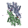
| |||||||||||||||||||||||||||
|---|---|---|---|---|---|---|---|---|---|---|---|---|---|---|---|---|---|---|---|---|---|---|---|---|---|---|---|---|
| 1 | 
| |||||||||||||||||||||||||||
| 2 | 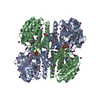
| |||||||||||||||||||||||||||
| Unit cell |
| |||||||||||||||||||||||||||
| Noncrystallographic symmetry (NCS) | NCS domain:
NCS domain segments:
|
-Components
-Protein , 1 types, 2 molecules AB
| #1: Protein | Mass: 43179.578 Da / Num. of mol.: 2 Source method: isolated from a genetically manipulated source Source: (gene. exp.) Mycobacterium smegmatis (bacteria) / Strain: MC2 155 / Gene: MSMEG_5491 / Plasmid: AVA0421 / Production host: |
|---|
-Non-polymers , 5 types, 701 molecules 


| #2: Chemical |  Mass: 22.990 Da / Num. of mol.: 2 / Source method: obtained synthetically / Formula: Na Mass: 22.990 Da / Num. of mol.: 2 / Source method: obtained synthetically / Formula: Na#3: Chemical | ChemComp-IOD /  Mass: 126.904 Da / Num. of mol.: 30 / Source method: obtained synthetically / Formula: I Mass: 126.904 Da / Num. of mol.: 30 / Source method: obtained synthetically / Formula: I#4: Chemical |  Mass: 62.068 Da / Num. of mol.: 2 / Source method: obtained synthetically / Formula: C2H6O2 Mass: 62.068 Da / Num. of mol.: 2 / Source method: obtained synthetically / Formula: C2H6O2#5: Chemical |  Mass: 787.566 Da / Num. of mol.: 2 / Source method: obtained synthetically / Formula: C27H35N9O15P2 Mass: 787.566 Da / Num. of mol.: 2 / Source method: obtained synthetically / Formula: C27H35N9O15P2#6: Water | ChemComp-HOH / | Mass: 18.015 Da / Num. of mol.: 665 / Source method: isolated from a natural source / Formula: H2O |
|---|
-Experimental details
-Experiment
| Experiment | Method: X-RAY DIFFRACTION / Number of used crystals: 1 |
|---|
- Sample preparation
| Crystal | Density Matthews: 2.34 Å3/Da / Density % sol: 47.5 % |
|---|---|
| Crystal grow | Temperature: 290 K / Method: vapor diffusion, sitting drop / pH: 8.5 Details: MD PACT SCEEN H10: 20MM NA/K PHOSPHATE, 100M BISTRISPROPANE pH 8.5, 20% PEG 3350, MYSMA.00247.B.A1 PW28486 AT 27MG/ML, SOAKED IN 50MM NA/K PHOSPHATE PH 7.0, 25% PEG 3350, 15% ETHYLENE ...Details: MD PACT SCEEN H10: 20MM NA/K PHOSPHATE, 100M BISTRISPROPANE pH 8.5, 20% PEG 3350, MYSMA.00247.B.A1 PW28486 AT 27MG/ML, SOAKED IN 50MM NA/K PHOSPHATE PH 7.0, 25% PEG 3350, 15% ETHYLENE GLYCOL, 500MM NAI, VAPOR DIFFUSION, SITTING DROP, TEMPERATURE 290K |
-Data collection
| Diffraction | Mean temperature: 100 K |
|---|---|
| Diffraction source | Source: ROTATING ANODE / Type: RIGAKU FR-E+ SUPERBRIGHT / Wavelength: 1.5418 Å |
| Detector | Type: RIGAKU SATURN 944+ / Detector: CCD / Date: Jun 22, 2009 |
| Radiation | Protocol: SINGLE WAVELENGTH / Monochromatic (M) / Laue (L): M / Scattering type: x-ray |
| Radiation wavelength | Wavelength: 1.5418 Å / Relative weight: 1 |
| Reflection | Resolution: 2.1→50 Å / Num. all: 46886 / Num. obs: 46378 / % possible obs: 98.9 % / Observed criterion σ(F): 0 / Observed criterion σ(I): -3 / Redundancy: 7.1 % / Biso Wilson estimate: 21.79 Å2 / Rmerge(I) obs: 0.045 / Net I/σ(I): 31.92 |
| Reflection shell | Resolution: 2.1→2.15 Å / Redundancy: 5.8 % / Rmerge(I) obs: 0.11 / Mean I/σ(I) obs: 14.6 / Num. unique all: 3278 / % possible all: 94 |
- Processing
| Software |
| ||||||||||||||||||||||||||||||||||||||||||||||||||||||||||||||||||||||||||||||||||||||||||||||||||||||||||||||||||||||||||||||||||||||||||||||||||||||||||||||||||||||||||
|---|---|---|---|---|---|---|---|---|---|---|---|---|---|---|---|---|---|---|---|---|---|---|---|---|---|---|---|---|---|---|---|---|---|---|---|---|---|---|---|---|---|---|---|---|---|---|---|---|---|---|---|---|---|---|---|---|---|---|---|---|---|---|---|---|---|---|---|---|---|---|---|---|---|---|---|---|---|---|---|---|---|---|---|---|---|---|---|---|---|---|---|---|---|---|---|---|---|---|---|---|---|---|---|---|---|---|---|---|---|---|---|---|---|---|---|---|---|---|---|---|---|---|---|---|---|---|---|---|---|---|---|---|---|---|---|---|---|---|---|---|---|---|---|---|---|---|---|---|---|---|---|---|---|---|---|---|---|---|---|---|---|---|---|---|---|---|---|---|---|---|---|
| Refinement | Method to determine structure: SAD / Resolution: 2.1→40.12 Å / Cor.coef. Fo:Fc: 0.953 / Cor.coef. Fo:Fc free: 0.93 / SU B: 6.856 / SU ML: 0.093 / Isotropic thermal model: isotropic, TLS / Cross valid method: THROUGHOUT / σ(F): 0 / ESU R Free: 0.166 / Stereochemistry target values: MAXIMUM LIKELIHOOD / Details: HYDROGENS HAVE BEEN ADDED IN THE
| ||||||||||||||||||||||||||||||||||||||||||||||||||||||||||||||||||||||||||||||||||||||||||||||||||||||||||||||||||||||||||||||||||||||||||||||||||||||||||||||||||||||||||
| Solvent computation | Ion probe radii: 0.8 Å / Shrinkage radii: 0.8 Å / VDW probe radii: 1.4 Å / Solvent model: MASK | ||||||||||||||||||||||||||||||||||||||||||||||||||||||||||||||||||||||||||||||||||||||||||||||||||||||||||||||||||||||||||||||||||||||||||||||||||||||||||||||||||||||||||
| Displacement parameters | Biso mean: 12.88 Å2
| ||||||||||||||||||||||||||||||||||||||||||||||||||||||||||||||||||||||||||||||||||||||||||||||||||||||||||||||||||||||||||||||||||||||||||||||||||||||||||||||||||||||||||
| Refinement step | Cycle: LAST / Resolution: 2.1→40.12 Å
| ||||||||||||||||||||||||||||||||||||||||||||||||||||||||||||||||||||||||||||||||||||||||||||||||||||||||||||||||||||||||||||||||||||||||||||||||||||||||||||||||||||||||||
| Refine LS restraints |
| ||||||||||||||||||||||||||||||||||||||||||||||||||||||||||||||||||||||||||||||||||||||||||||||||||||||||||||||||||||||||||||||||||||||||||||||||||||||||||||||||||||||||||
| Refine LS restraints NCS | Dom-ID: 1 / Auth asym-ID: A / Ens-ID: 1 / Number: 4772 / Refine-ID: X-RAY DIFFRACTION
| ||||||||||||||||||||||||||||||||||||||||||||||||||||||||||||||||||||||||||||||||||||||||||||||||||||||||||||||||||||||||||||||||||||||||||||||||||||||||||||||||||||||||||
| LS refinement shell | Resolution: 2.1→2.15 Å / Total num. of bins used: 20
| ||||||||||||||||||||||||||||||||||||||||||||||||||||||||||||||||||||||||||||||||||||||||||||||||||||||||||||||||||||||||||||||||||||||||||||||||||||||||||||||||||||||||||
| Refinement TLS params. | Method: refined / Refine-ID: X-RAY DIFFRACTION
| ||||||||||||||||||||||||||||||||||||||||||||||||||||||||||||||||||||||||||||||||||||||||||||||||||||||||||||||||||||||||||||||||||||||||||||||||||||||||||||||||||||||||||
| Refinement TLS group |
|
