 Movie
Movie Controller

[English] 日本語
 Yorodumi
- PDB-3iq6: Crystal structure of a tetrameric Zn-bound cytochrome cb562 compl... -
+ Open data

- Basic information
| Entry | Database: PDB / ID: 3iq6 | ||||||
|---|---|---|---|---|---|---|---|
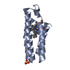
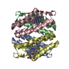
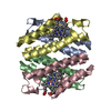
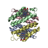
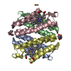
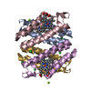
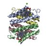
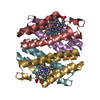
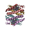
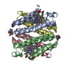
| Title | Crystal structure of a tetrameric Zn-bound cytochrome cb562 complex with covalently and non-covalently stabilized interfaces | ||||||
 Components Components | Soluble cytochrome b562 | ||||||
 Keywords Keywords | ELECTRON TRANSPORT / Tetramer of Four-Helix Bundles with Interfacial Disulfide Bonds / Heme / Iron / Metal-binding / Transport | ||||||
| Function / homology |  Function and homology information Function and homology informationelectron transport chain / periplasmic space / electron transfer activity / iron ion binding / heme binding Similarity search - Function | ||||||
| Biological species |  | ||||||
| Method |  X-RAY DIFFRACTION / SYNCHROTRON / MOLECULAR REPLACEMENT / molecular replacement / Resolution: 2.35 Å X-RAY DIFFRACTION / SYNCHROTRON / MOLECULAR REPLACEMENT / molecular replacement / Resolution: 2.35 Å | ||||||
 Authors Authors | Brodin, J.N. / Tezcan, F.A. | ||||||
 Citation Citation | Journal: J.Am.Chem.Soc. / Year: 2010 Title: Evolution of metal selectivity in templated protein interfaces. Authors: Brodin, J.D. / Medina-Morales, A. / Ni, T. / Salgado, E.N. / Ambroggio, X.I. / Tezcan, F.A. | ||||||
| History |
|
- Structure visualization
| Structure viewer | Molecule: MolmilJmol/JSmol |
|---|
- Downloads & links
-Download
| PDBx/mmCIF format | 3iq6.cif.gz | 184.9 KB | Display | PDBx/mmCIF format |
|---|---|---|---|---|
| PDB format | pdb3iq6.ent.gz | 150.6 KB | Display | PDB format |
| PDBx/mmJSON format | 3iq6.json.gz | Tree view | PDBx/mmJSON format | |
| Others |  Other downloads Other downloads |
-Validation report
| Arichive directory | https://data.pdbj.org/pub/pdb/validation_reports/iq/3iq6ftp://data.pdbj.org/pub/pdb/validation_reports/iq/3iq6 | HTTPS FTP |
|---|
-Related structure data
| Related structure data | 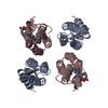 3iq5C  3m79C 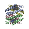 3hniS C: citing same article ( S: Starting model for refinement |
|---|---|
| Similar structure data |


-Links
 PDBj
PDBj


- Assembly
| Deposited unit | 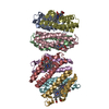
| ||||||||
|---|---|---|---|---|---|---|---|---|---|
| 1 | 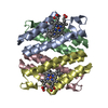
| ||||||||
| 2 | 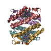
| ||||||||
| Unit cell |
| ||||||||
| Noncrystallographic symmetry (NCS) | NCS domain: (Details: chain A,B,C,D,E,F,G,H, using restrain) |
-Components
| #1: Protein | Mass: 11701.123 Da / Num. of mol.: 8 Mutation: R34A, L38A, Q41W, K42S, K59H, D66W, V69I, D73H, K77H, T96C, R98C, Y101C Source method: isolated from a natural source / Source: (natural) #2: Chemical | ChemComp-HEM /   Mass: 616.487 Da / Num. of mol.: 8 / Source method: obtained synthetically / Formula: C34H32FeN4O4 Mass: 616.487 Da / Num. of mol.: 8 / Source method: obtained synthetically / Formula: C34H32FeN4O4#3: Chemical | ChemComp-ZN /   Mass: 65.409 Da / Num. of mol.: 8 / Source method: obtained synthetically / Formula: Zn Mass: 65.409 Da / Num. of mol.: 8 / Source method: obtained synthetically / Formula: Zn#4: Water | ChemComp-HOH / |  Mass: 18.015 Da / Num. of mol.: 145 / Source method: isolated from a natural source / Formula: H2O Mass: 18.015 Da / Num. of mol.: 145 / Source method: isolated from a natural source / Formula: H2OHas protein modification | Y | |
|---|
-Experimental details
-Experiment
| Experiment | Method: X-RAY DIFFRACTION / Number of used crystals: 1 |
|---|
- Sample preparation
| Crystal | Density Matthews: 2.3 Å3/Da / Density % sol: 46.6 % |
|---|---|
| Crystal grow | Temperature: 293 K / Method: vapor diffusion, sitting drop / pH: 7.5 Details: 20% PEG 2000, 0.1M HEPES, 0.15 M Sodium chloride, 1.15 mM Zinc chloride, pH 7.5, VAPOR DIFFUSION, SITTING DROP, temperature 293K |
-Data collection
| Diffraction | Mean temperature: 100 K |
|---|---|
| Diffraction source | Source: SYNCHROTRON / Site: SSRL  / Beamline: BL9-2 / Wavelength: 0.98 Å / Beamline: BL9-2 / Wavelength: 0.98 Å |
| Detector | Type: MARMOSAIC 325 mm CCD / Detector: CCD / Date: May 16, 2009 Details: Flat collimating mirror, double crystal monochromator, toroid focusing mirror |
| Radiation | Monochromator: Double crystal / Protocol: SINGLE WAVELENGTH / Monochromatic (M) / Laue (L): M / Scattering type: x-ray |
| Radiation wavelength | Wavelength: 0.98 Å / Relative weight: 1 |
| Reflection | Resolution: 2.35→50 Å / Num. all: 36885 / Num. obs: 36701 / % possible obs: 99.5 % / Observed criterion σ(I): 2 / Redundancy: 4 % / Rmerge(I) obs: 0.098 / Rsym value: 0.098 / Net I/σ(I): 4.9 |
| Reflection shell | Resolution: 2.35→2.48 Å / Redundancy: 4 % / Rmerge(I) obs: 0.356 / Mean I/σ(I) obs: 1.8 / Num. unique all: 5295 / Rsym value: 0.356 / % possible all: 99.9 |
-Phasing
| Phasing | Method: molecular replacement |
|---|
- Processing
| Software |
| ||||||||||||||||||||||||||||||||||||||||||||||||||||||||
|---|---|---|---|---|---|---|---|---|---|---|---|---|---|---|---|---|---|---|---|---|---|---|---|---|---|---|---|---|---|---|---|---|---|---|---|---|---|---|---|---|---|---|---|---|---|---|---|---|---|---|---|---|---|---|---|---|---|
| Refinement | Method to determine structure: MOLECULAR REPLACEMENT Starting model: PDB entry 3HNI Resolution: 2.35→50 Å / Occupancy max: 1 / Occupancy min: 1 / FOM work R set: 0.763 / σ(F): 2 / σ(I): 2 / Stereochemistry target values: Engh & Huber
| ||||||||||||||||||||||||||||||||||||||||||||||||||||||||
| Solvent computation | Bsol: 39.728 Å2 | ||||||||||||||||||||||||||||||||||||||||||||||||||||||||
| Displacement parameters | Biso max: 97.37 Å2 / Biso mean: 46.922 Å2 / Biso min: 11.85 Å2
| ||||||||||||||||||||||||||||||||||||||||||||||||||||||||
| Refinement step | Cycle: LAST / Resolution: 2.35→50 Å
| ||||||||||||||||||||||||||||||||||||||||||||||||||||||||
| Refine LS restraints |
| ||||||||||||||||||||||||||||||||||||||||||||||||||||||||
| Refine LS restraints NCS |
| ||||||||||||||||||||||||||||||||||||||||||||||||||||||||
| LS refinement shell | Resolution: 2.35→2.37 Å
| ||||||||||||||||||||||||||||||||||||||||||||||||||||||||
| Xplor file |
|
