 Movie
Movie Controller

[English] 日本語
 Yorodumi
- PDB-3gvc: Crystal structure of probable short-chain dehydrogenase-reductase... -
+ Open data

- Basic information
| Entry | Database: PDB / ID: 3gvc | ||||||
|---|---|---|---|---|---|---|---|
| Title | Crystal structure of probable short-chain dehydrogenase-reductase from Mycobacterium tuberculosis | ||||||
 Components Components | Probable short-chain type dehydrogenase/reductase | ||||||
 Keywords Keywords | OXIDOREDUCTASE / SSGCID / deCODE / NIAID / UWPPG / SBRI / Structural Genomics / Seattle Structural Genomics Center for Infectious Disease | ||||||
| Function / homology |  Function and homology information Function and homology information | ||||||
| Biological species |   Mycobacterium tuberculosis (bacteria) Mycobacterium tuberculosis (bacteria) | ||||||
| Method |  X-RAY DIFFRACTION / SYNCHROTRON / MOLECULAR REPLACEMENT / molecular replacement / Resolution: 2.45 Å X-RAY DIFFRACTION / SYNCHROTRON / MOLECULAR REPLACEMENT / molecular replacement / Resolution: 2.45 Å | ||||||
 Authors Authors | Seattle Structural Genomics Center for Infectious Disease (SSGCID) | ||||||
 Citation Citation | Journal: Tuberculosis (Edinb) / Year: 2015 Title: Increasing the structural coverage of tuberculosis drug targets. Authors: Baugh, L. / Phan, I. / Begley, D.W. / Clifton, M.C. / Armour, B. / Dranow, D.M. / Taylor, B.M. / Muruthi, M.M. / Abendroth, J. / Fairman, J.W. / Fox, D. / Dieterich, S.H. / Staker, B.L. / ...Authors: Baugh, L. / Phan, I. / Begley, D.W. / Clifton, M.C. / Armour, B. / Dranow, D.M. / Taylor, B.M. / Muruthi, M.M. / Abendroth, J. / Fairman, J.W. / Fox, D. / Dieterich, S.H. / Staker, B.L. / Gardberg, A.S. / Choi, R. / Hewitt, S.N. / Napuli, A.J. / Myers, J. / Barrett, L.K. / Zhang, Y. / Ferrell, M. / Mundt, E. / Thompkins, K. / Tran, N. / Lyons-Abbott, S. / Abramov, A. / Sekar, A. / Serbzhinskiy, D. / Lorimer, D. / Buchko, G.W. / Stacy, R. / Stewart, L.J. / Edwards, T.E. / Van Voorhis, W.C. / Myler, P.J. | ||||||
| History |
|
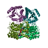
- Structure visualization
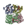
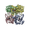
| Structure viewer | Molecule: MolmilJmol/JSmol |
|---|
- Downloads & links
-Download
| PDBx/mmCIF format | 3gvc.cif.gz | 185.2 KB | Display | PDBx/mmCIF format |
|---|---|---|---|---|
| PDB format | pdb3gvc.ent.gz | 147.2 KB | Display | PDB format |
| PDBx/mmJSON format | 3gvc.json.gz | Tree view | PDBx/mmJSON format | |
| Others |  Other downloads Other downloads |
-Validation report
| Arichive directory | https://data.pdbj.org/pub/pdb/validation_reports/gv/3gvcftp://data.pdbj.org/pub/pdb/validation_reports/gv/3gvc | HTTPS FTP |
|---|
-Related structure data
| Related structure data |  3gvgC  3gwcC  3h7fC  3h81C 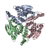 3he2C 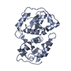 3hwiC 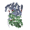 3hwkC 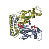 3hzgC  3icoC  3khpC  3llsC  3moyC 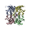 3mpzC 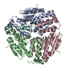 3mybC 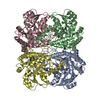 3ndnC 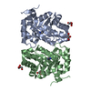 3ndoC 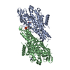 3nf4C  3ng3C 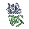 3njdC 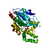 3nwoC 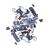 3o0mC 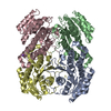 3o38C 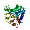 3oc6C 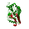 3oc7C 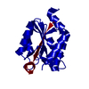 3oi9C 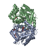 3oksC 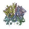 3omeC 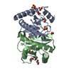 3p0tC 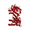 3p2yC 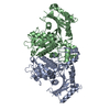 3p4iC 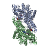 3p4tC 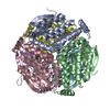 3p5mC 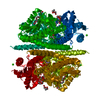 3p85C 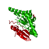 3pe8C  3pk0C 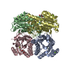 3ppiC 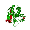 3pzyC 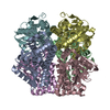 3q1tC 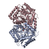 3q8nC 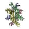 3qbpC 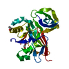 3qdfC 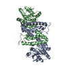 3qhaC 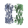 3qivC 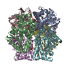 3qk8C 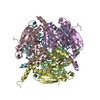 3qkaC  3qljC  3qmjC  3qreC  3quaC 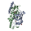 3quvC 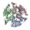 3qxiC 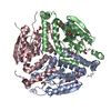 3qxzC 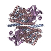 3qyrC 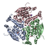 3r0oC 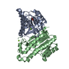 3r1iC 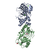 3r1jC 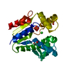 3r20C 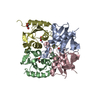 3r2nC 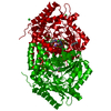 3r4tC  3r6hC  3r6oC 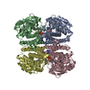 3r7kC 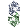 3r8cC 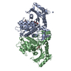 3r9pC 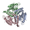 3r9qC 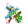 3r9rC 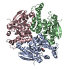 3r9sC 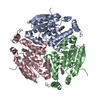 3r9tC 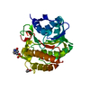 3rd5C 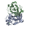 3rd7C 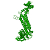 3rd8C 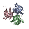 3rfqC 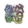 3rihC 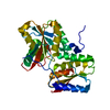 3rr2C 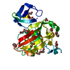 3rr6C 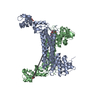 3rrpC 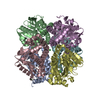 3rrvC 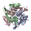 3rsiC 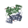 3rv2C 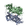 3s82C 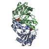 3sbxC  3sf6C 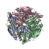 3sllC 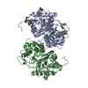 3svkC 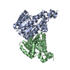 3svtC 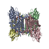 3swoC 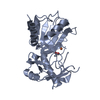 3swtC 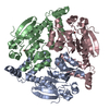 3swxC 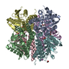 3t3wC 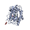 3tavC 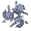 3tcrC 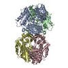 3tdeC 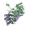 3tjrC 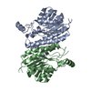 3tl3C 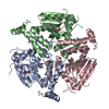 3tlfC  3trrC 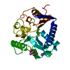 3tx2C 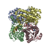 3tzqC 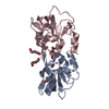 3tzuC 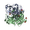 3u0aC 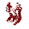 3ucxC 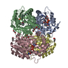 3uveC 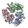 4di1C 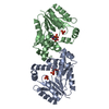 4dieC 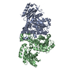 4dq8C 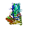 4dxlC 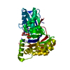 4ed4C  4egeC 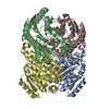 4egfC 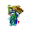 4emdC  4eo9C  4eyeC  4f3wC  4f47C 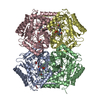 4ffcC 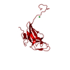 4gk6C 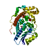 4hdtC 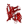 4hr3C 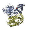 4i1yC 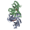 4ijnC 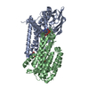 4iv6C 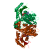 4iz9C 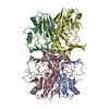 4j5iC 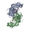 4kamC 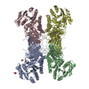 4lgvC 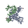 4o2dC  1iy8S S: Starting model for refinement C: citing same article ( |
|---|---|
| Similar structure data | |
| Other databases |
-Links
 PDBj
PDBj


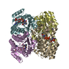
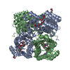
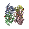
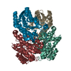
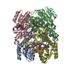
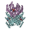
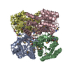
- Assembly
| Deposited unit | 
| ||||||||
|---|---|---|---|---|---|---|---|---|---|
| 1 | 
| ||||||||
| 2 | 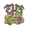
| ||||||||
| Unit cell |
|
-Components
| #1: Protein | Mass: 28146.250 Da / Num. of mol.: 4 Source method: isolated from a genetically manipulated source Details: Expressed with an N-terminal hexahis tag fused with a 3C protease substrate linker but not-cleaved prior to crystallization Source: (gene. exp.) Mycobacterium tuberculosis (bacteria) / Strain: H37Rv / Gene: MT1991, Rv1941 / Production host: References: UniProt: P95273, UniProt: I6XZC4*PLUS, Oxidoreductases #2: Water | ChemComp-HOH / |  Mass: 18.015 Da / Num. of mol.: 240 / Source method: isolated from a natural source / Formula: H2O Mass: 18.015 Da / Num. of mol.: 240 / Source method: isolated from a natural source / Formula: H2O |
|---|
-Experimental details
-Experiment
| Experiment | Method: X-RAY DIFFRACTION / Number of used crystals: 1 |
|---|
- Sample preparation
| Crystal | Density Matthews: 2.21 Å3/Da / Density % sol: 44.3 % |
|---|---|
| Crystal grow | Temperature: 289 K / Method: vapor diffusion, sitting drop / pH: 7 Details: Emerald Wizard Full screen condition F6, 20% PEG 3000, 0.1 M Tris, 0.2 M CaCl2, 11.8 mg/mL protein, crystal ID 202293f6, 25% glycerol as cryo-protectant, pH 7.0, VAPOR DIFFUSION, SITTING DROP, temperature 289K |
-Data collection
| Diffraction | Mean temperature: 100 K |
|---|---|
| Diffraction source | Source: SYNCHROTRON / Site: APS  / Beamline: 23-ID-D / Wavelength: 0.97933 Å / Beamline: 23-ID-D / Wavelength: 0.97933 Å |
| Detector | Date: Mar 19, 2009 |
| Radiation | Protocol: SINGLE WAVELENGTH / Monochromatic (M) / Laue (L): M / Scattering type: x-ray |
| Radiation wavelength | Wavelength: 0.97933 Å / Relative weight: 1 |
| Reflection | Resolution: 2.45→20 Å / Num. obs: 36246 / % possible obs: 98.8 % / Observed criterion σ(I): -3 / Biso Wilson estimate: 32.734 Å2 / Rmerge(I) obs: 0.135 / Net I/σ(I): 7.97 |
| Reflection shell | Resolution: 2.45→2.51 Å / Rmerge(I) obs: 0.561 / Mean I/σ(I) obs: 2.2 / Num. measured obs: 7568 / Num. unique all: 2654 / Num. unique obs: 2654 / % possible all: 99.5 |
-Phasing
| Phasing | Method: molecular replacement |
|---|
- Processing
| Software |
| |||||||||||||||||||||||||||||||||||||||||||||||||||||||||||||||||||||||||||||||||||||
|---|---|---|---|---|---|---|---|---|---|---|---|---|---|---|---|---|---|---|---|---|---|---|---|---|---|---|---|---|---|---|---|---|---|---|---|---|---|---|---|---|---|---|---|---|---|---|---|---|---|---|---|---|---|---|---|---|---|---|---|---|---|---|---|---|---|---|---|---|---|---|---|---|---|---|---|---|---|---|---|---|---|---|---|---|---|---|
| Refinement | Method to determine structure: MOLECULAR REPLACEMENT Starting model: 1IY8 Resolution: 2.45→19.9 Å / Cor.coef. Fo:Fc: 0.939 / Cor.coef. Fo:Fc free: 0.897 / WRfactor Rfree: 0.213 / WRfactor Rwork: 0.166 / Occupancy max: 1 / Occupancy min: 1 / FOM work R set: 0.83 / SU B: 9.386 / SU ML: 0.211 / SU R Cruickshank DPI: 0.549 / SU Rfree: 0.282 / Cross valid method: THROUGHOUT / σ(F): 0 / ESU R: 0.548 / ESU R Free: 0.283 / Stereochemistry target values: MAXIMUM LIKELIHOOD Details: HYDROGENS HAVE BEEN ADDED IN THE RIDING POSITIONS. U VALUES: REFINED INDIVIDUALLY
| |||||||||||||||||||||||||||||||||||||||||||||||||||||||||||||||||||||||||||||||||||||
| Solvent computation | Ion probe radii: 0.8 Å / Shrinkage radii: 0.8 Å / VDW probe radii: 1.4 Å / Solvent model: MASK | |||||||||||||||||||||||||||||||||||||||||||||||||||||||||||||||||||||||||||||||||||||
| Displacement parameters | Biso max: 74.25 Å2 / Biso mean: 25.165 Å2 / Biso min: 4.34 Å2
| |||||||||||||||||||||||||||||||||||||||||||||||||||||||||||||||||||||||||||||||||||||
| Refinement step | Cycle: LAST / Resolution: 2.45→19.9 Å
| |||||||||||||||||||||||||||||||||||||||||||||||||||||||||||||||||||||||||||||||||||||
| Refine LS restraints |
| |||||||||||||||||||||||||||||||||||||||||||||||||||||||||||||||||||||||||||||||||||||
| LS refinement shell | Resolution: 2.45→2.514 Å / Total num. of bins used: 20
|
