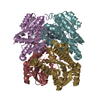
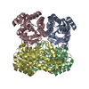
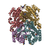
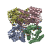
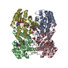
| Deposited unit | A: Retinal dehydrogenase/reductase 3
B: Retinal dehydrogenase/reductase 3
C: Retinal dehydrogenase/reductase 3
D: Retinal dehydrogenase/reductase 3
E: Retinal dehydrogenase/reductase 3
F: Retinal dehydrogenase/reductase 3
G: Retinal dehydrogenase/reductase 3
H: Retinal dehydrogenase/reductase 3
I: Retinal dehydrogenase/reductase 3
J: Retinal dehydrogenase/reductase 3
K: Retinal dehydrogenase/reductase 3
L: Retinal dehydrogenase/reductase 3
M: Retinal dehydrogenase/reductase 3
N: Retinal dehydrogenase/reductase 3
O: Retinal dehydrogenase/reductase 3
P: Retinal dehydrogenase/reductase 3
| Theoretical mass | Number of molelcules |
|---|
| Total (without water) | 453,270 | 16 |
|---|
| Polymers | 453,270 | 16 |
|---|
| Non-polymers | 0 | 0 |
|---|
| Water | 22,537 | 1251 |
|---|
|
|---|
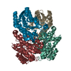
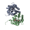
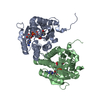
| 1 | A: Retinal dehydrogenase/reductase 3
B: Retinal dehydrogenase/reductase 3
C: Retinal dehydrogenase/reductase 3
D: Retinal dehydrogenase/reductase 3
| Theoretical mass | Number of molelcules |
|---|
| Total (without water) | 113,318 | 4 |
|---|
| Polymers | 113,318 | 4 |
|---|
| Non-polymers | 0 | 0 |
|---|
| Water | 72 | 4 |
|---|
| Type | Name | Symmetry operation | Number |
|---|
| identity operation | 1_555 | x,y,z | 1 |
| Buried area | 13490 Å2 |
|---|
| ΔGint | -104 kcal/mol |
|---|
| Surface area | 34930 Å2 |
|---|
| Method | PISA |
|---|
|
|---|
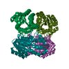
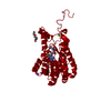
| 2 | E: Retinal dehydrogenase/reductase 3
F: Retinal dehydrogenase/reductase 3
G: Retinal dehydrogenase/reductase 3
H: Retinal dehydrogenase/reductase 3
| Theoretical mass | Number of molelcules |
|---|
| Total (without water) | 113,318 | 4 |
|---|
| Polymers | 113,318 | 4 |
|---|
| Non-polymers | 0 | 0 |
|---|
| Water | 72 | 4 |
|---|
| Type | Name | Symmetry operation | Number |
|---|
| identity operation | 1_555 | x,y,z | 1 |
| Buried area | 13470 Å2 |
|---|
| ΔGint | -109 kcal/mol |
|---|
| Surface area | 35220 Å2 |
|---|
| Method | PISA |
|---|
|
|---|
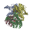
| 3 | I: Retinal dehydrogenase/reductase 3
J: Retinal dehydrogenase/reductase 3
K: Retinal dehydrogenase/reductase 3
L: Retinal dehydrogenase/reductase 3
| Theoretical mass | Number of molelcules |
|---|
| Total (without water) | 113,318 | 4 |
|---|
| Polymers | 113,318 | 4 |
|---|
| Non-polymers | 0 | 0 |
|---|
| Water | 72 | 4 |
|---|
| Type | Name | Symmetry operation | Number |
|---|
| identity operation | 1_555 | x,y,z | 1 |
| Buried area | 13330 Å2 |
|---|
| ΔGint | -107 kcal/mol |
|---|
| Surface area | 35220 Å2 |
|---|
| Method | PISA |
|---|
|
|---|
| 4 | M: Retinal dehydrogenase/reductase 3
N: Retinal dehydrogenase/reductase 3
O: Retinal dehydrogenase/reductase 3
P: Retinal dehydrogenase/reductase 3
| Theoretical mass | Number of molelcules |
|---|
| Total (without water) | 113,318 | 4 |
|---|
| Polymers | 113,318 | 4 |
|---|
| Non-polymers | 0 | 0 |
|---|
| Water | 72 | 4 |
|---|
| Type | Name | Symmetry operation | Number |
|---|
| identity operation | 1_555 | x,y,z | 1 |
| Buried area | 13160 Å2 |
|---|
| ΔGint | -112 kcal/mol |
|---|
| Surface area | 35460 Å2 |
|---|
| Method | PISA |
|---|
|
|---|
| Unit cell | | Length a, b, c (Å) | 167.115, 98.823, 167.463 |
|---|
| Angle α, β, γ (deg.) | 90.00, 115.87, 90.00 |
|---|
| Int Tables number | 4 |
|---|
| Space group name H-M | P1211 |
|---|
|
|---|
| Noncrystallographic symmetry (NCS) | NCS domain: | ID | Ens-ID | Details (eV) |
|---|
| 1 | 1 | A| 2 | 1 | C| 3 | 1 | E| 4 | 1 | G| 5 | 1 | I| 6 | 1 | K| 7 | 1 | M| 8 | 1 | O| 1 | 2 | D| 2 | 2 | B| 3 | 2 | F| 4 | 2 | H| 5 | 2 | J| 6 | 2 | L| 7 | 2 | N| 8 | 2 | P | | | | | | | | | | | | | | | |
NCS domain segments: Component-ID: 1 / Refine code: 2 | Dom-ID | Ens-ID | Beg auth comp-ID | Beg label comp-ID | End auth comp-ID | End label comp-ID | Auth asym-ID | Label asym-ID | Auth seq-ID | Label seq-ID |
|---|
| 1 | 1 | GLYGLYTYRTYRAA| 4 - 253 | 4 - 253 | | 2 | 1 | GLYGLYTYRTYRCC| 4 - 253 | 4 - 253 | | 3 | 1 | GLYGLYTYRTYREE| 4 - 253 | 4 - 253 | | 4 | 1 | ARGARGGLYGLYGG| 6 - 252 | 6 - 252 | | 5 | 1 | TYRTYRTYRTYRII| 7 - 253 | 7 - 253 | | 6 | 1 | GLYGLYTYRTYRKK| 4 - 253 | 4 - 253 | | 7 | 1 | LYSLYSTYRTYRMM| 10 - 253 | 10 - 253 | | 8 | 1 | TYRTYRTYRTYROO| 7 - 253 | 7 - 253 | | 1 | 2 | GLYGLYSERSERDD| 4 - 258 | 4 - 258 | | 2 | 2 | THRTHRALAALABB| 3 - 257 | 3 - 257 | | 3 | 2 | GLYGLYARGARGFF| 4 - 259 | 4 - 259 | | 4 | 2 | THRTHRSERSERHH| 5 - 258 | 5 - 258 | | 5 | 2 | GLYGLYSERSERJ| J | | | | | | | | | | | | | | | | | | | | | | | | | | | | | | | | | | | | | | | | | | | | | | | | | | | | | | | | | | | | | | | | | | | | | | | | | | | | | |
|
|---|
 Movie
Movie Controller

 Yorodumi
 Open data
 Basic information
 Components
 Keywords
 Function and homology information
 Homo sapiens (human)
 X-RAY DIFFRACTION /
  Authors
 Citation
 Structure visualization
 Downloads & links
 Other downloads
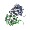

 PDBj
PDBj


 Assembly