 Movie
Movie Controller


+ Open data

- Basic information
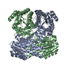
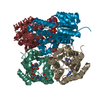
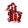
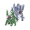
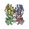
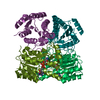
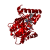
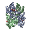
| Entry | Database: PDB / ID: 1geg | ||||||
|---|---|---|---|---|---|---|---|
| Title | CRYATAL STRUCTURE ANALYSIS OF MESO-2,3-BUTANEDIOL DEHYDROGENASE | ||||||
 Components Components | ACETOIN REDUCTASE | ||||||
 Keywords Keywords | OXIDOREDUCTASE / SDR FAMILY | ||||||
| Function / homology |  Function and homology information Function and homology informationdiacetyl reductase [(S)-acetoin forming] / diacetyl reductase ((S)-acetoin forming) (NAD+) activity / acetoin catabolic process Similarity search - Function | ||||||
| Biological species |  Klebsiella pneumoniae (bacteria) Klebsiella pneumoniae (bacteria) | ||||||
| Method |  X-RAY DIFFRACTION / SYNCHROTRON / MOLECULAR REPLACEMENT / Resolution: 1.7 Å X-RAY DIFFRACTION / SYNCHROTRON / MOLECULAR REPLACEMENT / Resolution: 1.7 Å | ||||||
 Authors Authors | Otagiri, M. / Kurisu, G. / Ui, S. / Kusunoki, M. | ||||||
 Citation Citation | Journal: J.Biochem. / Year: 2001 Title: Crystal structure of meso-2,3-butanediol dehydrogenase in a complex with NAD+ and inhibitor mercaptoethanol at 1.7 A resolution for understanding of chiral substrate recognition mechanisms. Authors: Otagiri, M. / Kurisu, G. / Ui, S. / Takusagawa, Y. / Ohkuma, M. / Kudo, T. / Kusunoki, M. | ||||||
| History |
|
- Structure visualization
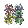
| Structure viewer | Molecule: MolmilJmol/JSmol |
|---|
- Downloads & links
-Download
| PDBx/mmCIF format | 1geg.cif.gz | 388.7 KB | Display | PDBx/mmCIF format |
|---|---|---|---|---|
| PDB format | pdb1geg.ent.gz | 319.6 KB | Display | PDB format |
| PDBx/mmJSON format | 1geg.json.gz | Tree view | PDBx/mmJSON format | |
| Others |  Other downloads Other downloads |
-Validation report
| Arichive directory | https://data.pdbj.org/pub/pdb/validation_reports/ge/1gegftp://data.pdbj.org/pub/pdb/validation_reports/ge/1geg | HTTPS FTP |
|---|
-Related structure data
| Similar structure data |
|---|

-Links
 PDBj
PDBj


- Assembly
| Deposited unit | 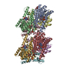
| ||||||||
|---|---|---|---|---|---|---|---|---|---|
| 1 | 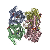
| ||||||||
| 2 | 
| ||||||||
| Unit cell |
|
-Components
-Protein / Sugars , 2 types, 16 molecules ABCDEFGH
| #1: Protein | Mass: 26618.299 Da / Num. of mol.: 8 Source method: isolated from a genetically manipulated source Source: (gene. exp.) Klebsiella pneumoniae (bacteria) / Plasmid: PUC119 / Production host: #2: Sugar | ChemComp-GLC /  Type: D-saccharide, alpha linking / Mass: 180.156 Da / Num. of mol.: 8 Type: D-saccharide, alpha linking / Mass: 180.156 Da / Num. of mol.: 8Source method: isolated from a genetically manipulated source Formula: C6H12O6 |
|---|
-Non-polymers , 4 types, 859 molecules 


| #3: Chemical | ChemComp-MG /  Mass: 24.305 Da / Num. of mol.: 4 / Source method: obtained synthetically / Formula: Mg Mass: 24.305 Da / Num. of mol.: 4 / Source method: obtained synthetically / Formula: Mg#4: Chemical | ChemComp-NAD /  Mass: 663.425 Da / Num. of mol.: 8 / Source method: obtained synthetically / Formula: C21H27N7O14P2 / Comment: NAD*YM Mass: 663.425 Da / Num. of mol.: 8 / Source method: obtained synthetically / Formula: C21H27N7O14P2 / Comment: NAD*YM#5: Chemical | ChemComp-BME /  Mass: 78.133 Da / Num. of mol.: 8 / Source method: obtained synthetically / Formula: C2H6OS Mass: 78.133 Da / Num. of mol.: 8 / Source method: obtained synthetically / Formula: C2H6OS#6: Water | ChemComp-HOH / | Mass: 18.015 Da / Num. of mol.: 839 / Source method: isolated from a natural source / Formula: H2O |
|---|
-Experimental details
-Experiment
| Experiment | Method: X-RAY DIFFRACTION / Number of used crystals: 1 |
|---|
- Sample preparation
| Crystal | Density Matthews: 2.22 Å3/Da / Density % sol: 44.5 % | ||||||||||||||||||||||||||||||||||||||||||||||||||||||||||||||||||
|---|---|---|---|---|---|---|---|---|---|---|---|---|---|---|---|---|---|---|---|---|---|---|---|---|---|---|---|---|---|---|---|---|---|---|---|---|---|---|---|---|---|---|---|---|---|---|---|---|---|---|---|---|---|---|---|---|---|---|---|---|---|---|---|---|---|---|---|
| Crystal grow | Temperature: 293 K / Method: vapor diffusion, hanging drop / pH: 7 Details: PEG 6000, 100mM HEPES, magnesium chloride, glucose, 2-mercaptoethanol, pH 7.0, VAPOR DIFFUSION, HANGING DROP, temperature 293K | ||||||||||||||||||||||||||||||||||||||||||||||||||||||||||||||||||
| Crystal grow | *PLUS pH: 8 | ||||||||||||||||||||||||||||||||||||||||||||||||||||||||||||||||||
| Components of the solutions | *PLUS
|
-Data collection
| Diffraction | Mean temperature: 100 K |
|---|---|
| Diffraction source | Source: SYNCHROTRON / Site: SPring-8  / Beamline: BL40B2 / Wavelength: 0.9 Å / Beamline: BL40B2 / Wavelength: 0.9 Å |
| Detector | Type: RIGAKU RAXIS IV++ / Detector: IMAGE PLATE / Date: Dec 15, 1999 |
| Radiation | Protocol: SINGLE WAVELENGTH / Monochromatic (M) / Laue (L): M / Scattering type: x-ray |
| Radiation wavelength | Wavelength: 0.9 Å / Relative weight: 1 |
| Reflection | Resolution: 1.7→40 Å / Num. all: 203543 / % possible obs: 100 % / Redundancy: 3.7 % / Rmerge(I) obs: 0.054 |
| Reflection shell | Resolution: 1.7→1.79 Å / Redundancy: 3.6 % / Rmerge(I) obs: 0.192 / % possible all: 100 |
| Reflection | *PLUS Lowest resolution: 40 Å / Num. obs: 203217 / % possible obs: 100 % |
| Reflection shell | *PLUS Highest resolution: 1.7 Å / % possible obs: 100 % |
- Processing
| Software |
| ||||||||||||||||||
|---|---|---|---|---|---|---|---|---|---|---|---|---|---|---|---|---|---|---|---|
| Refinement | Method to determine structure: MOLECULAR REPLACEMENT / Resolution: 1.7→40 Å / σ(F): 2
| ||||||||||||||||||
| Refinement step | Cycle: LAST / Resolution: 1.7→40 Å
| ||||||||||||||||||
| Refine LS restraints |
| ||||||||||||||||||
| Software | *PLUS Name: CNS / Version: 0.9 / Classification: refinement | ||||||||||||||||||
| Refinement | *PLUS Highest resolution: 1.7 Å / Lowest resolution: 40 Å / σ(F): 2 / Rfactor obs: 0.193 | ||||||||||||||||||
| Solvent computation | *PLUS | ||||||||||||||||||
| Displacement parameters | *PLUS | ||||||||||||||||||
| Refine LS restraints | *PLUS
|
