 Movie
Movie Controller

[English] 日本語
 Yorodumi
- PDB-1edo: THE X-RAY STRUCTURE OF BETA-KETO ACYL CARRIER PROTEIN REDUCTASE F... -
+ Open data

- Basic information
| Entry | Database: PDB / ID: 1edo | ||||||
|---|---|---|---|---|---|---|---|
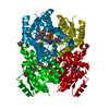
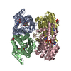
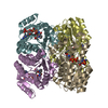
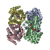
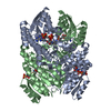
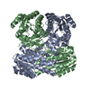
| Title | THE X-RAY STRUCTURE OF BETA-KETO ACYL CARRIER PROTEIN REDUCTASE FROM BRASSICA NAPUS COMPLEXED WITH NADP+ | ||||||
 Components Components | BETA-KETO ACYL CARRIER PROTEIN REDUCTASE | ||||||
 Keywords Keywords | OXIDOREDUCTASE / nucleotide fold / rossmann fold | ||||||
| Function / homology |  Function and homology information Function and homology information3-oxoacyl-[acyl-carrier-protein] reductase / 3-oxoacyl-[acyl-carrier-protein] reductase (NADPH) activity / chloroplast / NAD binding / fatty acid biosynthetic process Similarity search - Function | ||||||
| Biological species |  | ||||||
| Method |  X-RAY DIFFRACTION / Resolution: 2.3 Å X-RAY DIFFRACTION / Resolution: 2.3 Å | ||||||
 Authors Authors | Fisher, M. / Kroon, J.T. / Martindale, W. / Stuitje, A.R. / Slabas, A.R. / Rafferty, J.B. | ||||||
 Citation Citation | Journal: Structure Fold.Des. / Year: 2000 Title: The X-ray structure of Brassica napus beta-keto acyl carrier protein reductase and its implications for substrate binding and catalysis. Authors: Fisher, M. / Kroon, J.T. / Martindale, W. / Stuitje, A.R. / Slabas, A.R. / Rafferty, J.B. #1: Journal: Acta Crystallogr.,Sect.D / Year: 1999Title: Crystallization of the NADP-dependent beta-keto acyl-carrier protein reductase from Brassica napus Authors: Fisher, M. / Sedelnikova, S.E. / Martindale, W. / Simon, J.W. / Slabas, A.R. / Rafferty, J.B. | ||||||
| History |
|
- Structure visualization
| Structure viewer | Molecule: MolmilJmol/JSmol |
|---|
- Downloads & links
-Download
| PDBx/mmCIF format | 1edo.cif.gz | 60 KB | Display | PDBx/mmCIF format |
|---|---|---|---|---|
| PDB format | pdb1edo.ent.gz | 43.5 KB | Display | PDB format |
| PDBx/mmJSON format | 1edo.json.gz | Tree view | PDBx/mmJSON format | |
| Others |  Other downloads Other downloads |
-Validation report
| Arichive directory | https://data.pdbj.org/pub/pdb/validation_reports/ed/1edoftp://data.pdbj.org/pub/pdb/validation_reports/ed/1edo | HTTPS FTP |
|---|
-Related structure data
| Similar structure data |
|---|


-Links
 PDBj
PDBj


- Assembly
| Deposited unit | 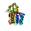
| ||||||||
|---|---|---|---|---|---|---|---|---|---|
| 1 | 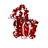
| ||||||||
| Unit cell |
| ||||||||
| Details | The biological assembly is a tetramer formed from the monomer and generated by 222 symmetry |
-Components
| #1: Protein | Mass: 25359.418 Da / Num. of mol.: 1 Source method: isolated from a genetically manipulated source Source: (gene. exp.)  |
|---|---|
| #2: Chemical | ChemComp-NAP /   Mass: 743.405 Da / Num. of mol.: 1 / Source method: obtained synthetically / Formula: C21H28N7O17P3 Mass: 743.405 Da / Num. of mol.: 1 / Source method: obtained synthetically / Formula: C21H28N7O17P3 |
| #3: Water | ChemComp-HOH /  Mass: 18.015 Da / Num. of mol.: 124 / Source method: isolated from a natural source / Formula: H2O Mass: 18.015 Da / Num. of mol.: 124 / Source method: isolated from a natural source / Formula: H2O |
-Experimental details
-Experiment
| Experiment | Method: X-RAY DIFFRACTION / Number of used crystals: 1 |
|---|
- Sample preparation
| Crystal | Density Matthews: 4.41 Å3/Da / Density % sol: 72.08 % | |||||||||||||||||||||||||||||||||||
|---|---|---|---|---|---|---|---|---|---|---|---|---|---|---|---|---|---|---|---|---|---|---|---|---|---|---|---|---|---|---|---|---|---|---|---|---|
| Crystal grow | Temperature: 290 K / Method: vapor diffusion, hanging drop / pH: 4 Details: PEG1500, Sodium citrate, pH 4, VAPOR DIFFUSION, HANGING DROP, temperature 290K | |||||||||||||||||||||||||||||||||||
| Crystal | *PLUS Density % sol: 73 % | |||||||||||||||||||||||||||||||||||
| Crystal grow | *PLUS Details: Fisher, M., (2000) Acta Crystallogr., Sect.D, 56, 86. | |||||||||||||||||||||||||||||||||||
| Components of the solutions | *PLUS
|
-Data collection
| Diffraction | Mean temperature: 298 K |
|---|---|
| Diffraction source | Source: ROTATING ANODE / Type: RIGAKU RU200 / Wavelength: 1.542 |
| Detector | Type: MARRESEARCH / Detector: IMAGE PLATE / Date: Oct 13, 1998 |
| Radiation | Protocol: SINGLE WAVELENGTH / Monochromatic (M) / Laue (L): M / Scattering type: x-ray |
| Radiation wavelength | Wavelength: 1.542 Å / Relative weight: 1 |
| Reflection | Resolution: 2.22→47.9 Å / Num. all: 102604 / Num. obs: 102604 / % possible obs: 95 % / Redundancy: 4.8 % / Rmerge(I) obs: 0.071 |
| Reflection shell | Resolution: 2.22→2.34 Å / Redundancy: 4.8 % / Rmerge(I) obs: 0.45 / Num. unique all: 21256 / % possible all: 91.5 |
| Reflection | *PLUS Num. obs: 21256 / % possible obs: 95 % / Num. measured all: 102604 |
| Reflection shell | *PLUS % possible obs: 91.5 % / Rmerge(I) obs: 0.45 |
- Processing
| Software |
| ||||||||||||||||||||
|---|---|---|---|---|---|---|---|---|---|---|---|---|---|---|---|---|---|---|---|---|---|
| Refinement | Resolution: 2.3→10 Å / Stereochemistry target values: Engh & Huber
| ||||||||||||||||||||
| Refinement step | Cycle: LAST / Resolution: 2.3→10 Å
| ||||||||||||||||||||
| Refine LS restraints |
| ||||||||||||||||||||
| Software | *PLUS Name: REFMAC / Classification: refinement | ||||||||||||||||||||
| Refinement | *PLUS Highest resolution: 2.3 Å / Lowest resolution: 10 Å / Rfactor obs: 0.19 / Rfactor Rwork: 0.19 | ||||||||||||||||||||
| Solvent computation | *PLUS | ||||||||||||||||||||
| Displacement parameters | *PLUS | ||||||||||||||||||||
| Refine LS restraints | *PLUS
|
