 Movie
Movie Controller

[English] 日本語
 Yorodumi
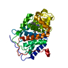
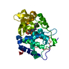
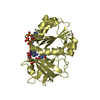
- PDB-5vml: Crystal Structure of Acetoacetyl-CoA Reductase from Burkholderia ... -
+ Open data

- Basic information
| Entry | Database: PDB / ID: 5vml | ||||||
|---|---|---|---|---|---|---|---|
| Title | Crystal Structure of Acetoacetyl-CoA Reductase from Burkholderia Pseudomallei 1710b with bound NADP | ||||||
 Components Components | Acetoacetyl-CoA reductase | ||||||
 Keywords Keywords | OXIDOREDUCTASE / SSGCID / Structural Genomics / Seattle Structural Genomics Center for Infectious Disease | ||||||
| Function / homology |  Function and homology information Function and homology informationacetoacetyl-CoA reductase / acetoacetyl-CoA reductase activity / poly-hydroxybutyrate biosynthetic process / monocarboxylic acid metabolic process / nucleotide binding / cytoplasm Similarity search - Function | ||||||
| Biological species |  Burkholderia pseudomallei (bacteria) Burkholderia pseudomallei (bacteria) | ||||||
| Method |  X-RAY DIFFRACTION / MOLECULAR REPLACEMENT / molecular replacement / Resolution: 1.7 Å X-RAY DIFFRACTION / MOLECULAR REPLACEMENT / molecular replacement / Resolution: 1.7 Å | ||||||
 Authors Authors | Seattle Structural Genomics Center for Infectious Disease (SSGCID) | ||||||
 Citation Citation | Journal: to be published Title: Crystal Structure of Acetoacetyl-CoA Reductase from Burkholderia Pseudomallei 1710b with bound NADP Authors: Dranow, D.M. / Conrady, D.G. / Lorimer, D.D. / Edwards, T.E. | ||||||
| History |
|
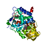
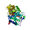
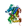
- Structure visualization
| Structure viewer | Molecule: MolmilJmol/JSmol |
|---|
- Downloads & links
-Download
| PDBx/mmCIF format | 5vml.cif.gz | 119.1 KB | Display | PDBx/mmCIF format |
|---|---|---|---|---|
| PDB format | pdb5vml.ent.gz | 88.6 KB | Display | PDB format |
| PDBx/mmJSON format | 5vml.json.gz | Tree view | PDBx/mmJSON format | |
| Others |  Other downloads Other downloads |
-Validation report
| Arichive directory | https://data.pdbj.org/pub/pdb/validation_reports/vm/5vmlftp://data.pdbj.org/pub/pdb/validation_reports/vm/5vml | HTTPS FTP |
|---|
-Related structure data
| Related structure data | 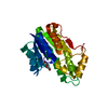 3ezlS S: Starting model for refinement |
|---|---|
| Similar structure data | |
| Other databases |


-Links
 PDBj
PDBj


- Assembly
| Deposited unit | 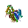
| ||||||||||||
|---|---|---|---|---|---|---|---|---|---|---|---|---|---|
| 1 |
| ||||||||||||
| 2 | 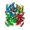
| ||||||||||||
| 3 | 
| ||||||||||||
| 4 | 
| ||||||||||||
| Unit cell |
| ||||||||||||
| Components on special symmetry positions |
| ||||||||||||
| Details | Monomer as determined by gel filtration. |
-Components
| #1: Protein | Mass: 29056.859 Da / Num. of mol.: 1 Source method: isolated from a genetically manipulated source Source: (gene. exp.) Burkholderia pseudomallei (strain 1710b) (bacteria)Strain: 1710b / Gene: phbB-1, BURPS1710b_2329 / Plasmid: BupsA.00010.e.A1 / Production host: |
|---|---|
| #2: Chemical | ChemComp-NAP /   Mass: 743.405 Da / Num. of mol.: 1 / Source method: obtained synthetically / Formula: C21H28N7O17P3 Mass: 743.405 Da / Num. of mol.: 1 / Source method: obtained synthetically / Formula: C21H28N7O17P3 |
| #3: Chemical | ChemComp-MPD / (  Mass: 118.174 Da / Num. of mol.: 1 / Source method: obtained synthetically / Formula: C6H14O2 / Comment: precipitant*YM Mass: 118.174 Da / Num. of mol.: 1 / Source method: obtained synthetically / Formula: C6H14O2 / Comment: precipitant*YM |
| #4: Water | ChemComp-HOH /  Mass: 18.015 Da / Num. of mol.: 282 / Source method: isolated from a natural source / Formula: H2O Mass: 18.015 Da / Num. of mol.: 282 / Source method: isolated from a natural source / Formula: H2O |
-Experimental details
-Experiment
| Experiment | Method: X-RAY DIFFRACTION / Number of used crystals: 1 |
|---|
- Sample preparation
| Crystal | Density Matthews: 2.18 Å3/Da / Density % sol: 43.58 % |
|---|---|
| Crystal grow | Temperature: 290 K / Method: vapor diffusion, sitting drop / pH: 7.5 Details: BupsA.00010.e.A1.PS00061 at 19.3 mg/ml incubated with 4 mM NADP, mixed 1:1 with Morpheus(c8): 12.5% (w/v) PEG-1000, 12.5% (w/v) PEG-3350, 12.5% (v/v) MPD, 0.1 M MOPS/ HEPES-Na, pH = 7.5, 0. ...Details: BupsA.00010.e.A1.PS00061 at 19.3 mg/ml incubated with 4 mM NADP, mixed 1:1 with Morpheus(c8): 12.5% (w/v) PEG-1000, 12.5% (w/v) PEG-3350, 12.5% (v/v) MPD, 0.1 M MOPS/ HEPES-Na, pH = 7.5, 0.03 M each sodium nitrate, disodium hydrogen phosphate, ammonium sulfate, crystals were soaked for 24 hours with 5 mM NADP in well solution, harvested directly |
-Data collection
| Diffraction | Mean temperature: 100 K |
|---|---|
| Diffraction source | Source: ROTATING ANODE / Type: RIGAKU FR-E+ SUPERBRIGHT / Wavelength: 1.5418 Å |
| Detector | Type: RIGAKU SATURN 944+ / Detector: CCD / Date: Mar 2, 2017 |
| Radiation | Monochromator: Rigaku Varimax HF / Protocol: SINGLE WAVELENGTH / Monochromatic (M) / Laue (L): M / Scattering type: x-ray |
| Radiation wavelength | Wavelength: 1.5418 Å / Relative weight: 1 |
| Reflection | Resolution: 1.7→50 Å / Num. obs: 28909 / % possible obs: 99.5 % / Redundancy: 8.9 % / Biso Wilson estimate: 15.87 Å2 / CC1/2: 0.999 / Rmerge(I) obs: 0.062 / Net I/σ(I): 22.03 |
| Reflection shell | Resolution: 1.7→1.74 Å / Redundancy: 4.3 % / Rmerge(I) obs: 0.458 / Num. unique obs: 1965 / CC1/2: 0.856 / % possible all: 93.2 |
-Phasing
| Phasing | Method: molecular replacement |
|---|
- Processing
| Software |
| |||||||||||||||||||||||||||||||||||||||||||||||||||||||||||||||||||||||||||||||||||||||||||||||||||||||||
|---|---|---|---|---|---|---|---|---|---|---|---|---|---|---|---|---|---|---|---|---|---|---|---|---|---|---|---|---|---|---|---|---|---|---|---|---|---|---|---|---|---|---|---|---|---|---|---|---|---|---|---|---|---|---|---|---|---|---|---|---|---|---|---|---|---|---|---|---|---|---|---|---|---|---|---|---|---|---|---|---|---|---|---|---|---|---|---|---|---|---|---|---|---|---|---|---|---|---|---|---|---|---|---|---|---|---|
| Refinement | Method to determine structure: MOLECULAR REPLACEMENT Starting model: 3EZL Resolution: 1.7→46.888 Å / SU ML: 0.16 / Cross valid method: FREE R-VALUE / σ(F): 1.35 / Phase error: 15.81
| |||||||||||||||||||||||||||||||||||||||||||||||||||||||||||||||||||||||||||||||||||||||||||||||||||||||||
| Solvent computation | Shrinkage radii: 0.9 Å / VDW probe radii: 1.11 Å | |||||||||||||||||||||||||||||||||||||||||||||||||||||||||||||||||||||||||||||||||||||||||||||||||||||||||
| Displacement parameters | Biso max: 73.31 Å2 / Biso mean: 19.0649 Å2 / Biso min: 7.89 Å2 | |||||||||||||||||||||||||||||||||||||||||||||||||||||||||||||||||||||||||||||||||||||||||||||||||||||||||
| Refinement step | Cycle: final / Resolution: 1.7→46.888 Å
| |||||||||||||||||||||||||||||||||||||||||||||||||||||||||||||||||||||||||||||||||||||||||||||||||||||||||
| Refine LS restraints |
| |||||||||||||||||||||||||||||||||||||||||||||||||||||||||||||||||||||||||||||||||||||||||||||||||||||||||
| LS refinement shell | Refine-ID: X-RAY DIFFRACTION / Rfactor Rfree error: 0 / Total num. of bins used: 14
| |||||||||||||||||||||||||||||||||||||||||||||||||||||||||||||||||||||||||||||||||||||||||||||||||||||||||
| Refinement TLS params. | Method: refined / Refine-ID: X-RAY DIFFRACTION
| |||||||||||||||||||||||||||||||||||||||||||||||||||||||||||||||||||||||||||||||||||||||||||||||||||||||||
| Refinement TLS group |
|
