 Movie
Movie Controller

[English] 日本語
 Yorodumi
- PDB-3ezl: Crystal Structure of Acetoacetyl-CoA Reductase from Burkholderia ... -
+ Open data

- Basic information
| Entry | Database: PDB / ID: 3ezl | ||||||
|---|---|---|---|---|---|---|---|
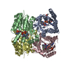
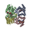
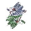
| Title | Crystal Structure of Acetoacetyl-CoA Reductase from Burkholderia Pseudomallei 1710b | ||||||
 Components Components | Acetoacetyl-CoA reductase | ||||||
 Keywords Keywords | OXIDOREDUCTASE / SSGCID / ACETOACETYL-COA REDUCTASE / BURKHOLDERIA PSEUDOMALLEI / Structural Genomics / Seattle Structural Genomics Center for Infectious Disease | ||||||
| Function / homology |  Function and homology information Function and homology informationacetoacetyl-CoA reductase / acetoacetyl-CoA reductase activity / poly-hydroxybutyrate biosynthetic process / monocarboxylic acid metabolic process / nucleotide binding / cytoplasm Similarity search - Function | ||||||
| Biological species |  Burkholderia pseudomallei 1710b (bacteria) Burkholderia pseudomallei 1710b (bacteria) | ||||||
| Method |  X-RAY DIFFRACTION / MOLECULAR REPLACEMENT / Resolution: 2.25 Å X-RAY DIFFRACTION / MOLECULAR REPLACEMENT / Resolution: 2.25 Å | ||||||
 Authors Authors | Seattle Structural Genomics Center for Infectious Disease (SSGCID) | ||||||
 Citation Citation | Journal: To be Published Title: Crystal Structure of Acetoacetyl-CoA Reductase from Burkholderia Pseudomallei 1710b Authors: Seattle Structural Genomics Center for Infectious Disease (SSGCID) | ||||||
| History |
|
- Structure visualization
| Structure viewer | Molecule: MolmilJmol/JSmol |
|---|
- Downloads & links
-Download
| PDBx/mmCIF format | 3ezl.cif.gz | 62.8 KB | Display | PDBx/mmCIF format |
|---|---|---|---|---|
| PDB format | pdb3ezl.ent.gz | 44.2 KB | Display | PDB format |
| PDBx/mmJSON format | 3ezl.json.gz | Tree view | PDBx/mmJSON format | |
| Others |  Other downloads Other downloads |
-Validation report
| Arichive directory | https://data.pdbj.org/pub/pdb/validation_reports/ez/3ezlftp://data.pdbj.org/pub/pdb/validation_reports/ez/3ezl | HTTPS FTP |
|---|
-Related structure data
| Related structure data |  1i01S S: Starting model for refinement |
|---|---|
| Similar structure data | |
| Other databases |


-Links
 PDBj
PDBj


- Assembly
| Deposited unit | 
| ||||||||
|---|---|---|---|---|---|---|---|---|---|
| 1 | 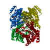
| ||||||||
| 2 |
| ||||||||
| Unit cell |
|
-Components
| #1: Protein | Mass: 27798.504 Da / Num. of mol.: 1 Source method: isolated from a genetically manipulated source Source: (gene. exp.) Burkholderia pseudomallei 1710b (bacteria)Strain: 1710B / Gene: phbB-1, BURPS1710b_2329 / Plasmid: AVA0421 / Production host: |
|---|---|
| #2: Chemical | ChemComp-P4C /   Mass: 324.367 Da / Num. of mol.: 1 / Source method: obtained synthetically / Formula: C14H28O8 Mass: 324.367 Da / Num. of mol.: 1 / Source method: obtained synthetically / Formula: C14H28O8 |
| #3: Water | ChemComp-HOH /  Mass: 18.015 Da / Num. of mol.: 136 / Source method: isolated from a natural source / Formula: H2O Mass: 18.015 Da / Num. of mol.: 136 / Source method: isolated from a natural source / Formula: H2O |
-Experimental details
-Experiment
| Experiment | Method: X-RAY DIFFRACTION / Number of used crystals: 1 |
|---|
- Sample preparation
| Crystal | Density Matthews: 2.24 Å3/Da / Density % sol: 45.13 % |
|---|---|
| Crystal grow | Temperature: 290 K / Method: vapor diffusion, sitting drop / pH: 7.5 Details: PROPLEX-96 A-1: 100MM HEPES PH 7.5, 20% PEG MME 2000, VAPOR DIFFUSION, SITTING DROP, temperature 290K, pH 7.50 |
-Data collection
| Diffraction | Mean temperature: 100 K |
|---|---|
| Diffraction source | Source: ROTATING ANODE / Type: RIGAKU MICROMAX-007 HF / Wavelength: 1.5418 / Wavelength: 1.5418 Å |
| Detector | Type: RIGAKU SATURN 944 / Detector: CCD / Date: Sep 8, 2008 |
| Radiation | Protocol: SINGLE WAVELENGTH / Monochromatic (M) / Laue (L): M / Scattering type: x-ray |
| Radiation wavelength | Wavelength: 1.5418 Å / Relative weight: 1 |
| Reflection | Resolution: 2.25→50 Å / Num. all: 12901 / Num. obs: 12901 / % possible obs: 98.3 % / Observed criterion σ(F): 0 / Observed criterion σ(I): -3 / Redundancy: 12.9 % / Biso Wilson estimate: 32.87 Å2 / Rmerge(I) obs: 0.087 / Rsym value: 0.087 |
| Reflection shell | Resolution: 2.25→2.31 Å / Rmerge(I) obs: 0.448 / Mean I/σ(I) obs: 4.9 / % possible all: 87.2 |
- Processing
| Software |
| |||||||||||||||||||||||||||||||||||||||||||||||||||||||||||||||||||||||||||||||||||||
|---|---|---|---|---|---|---|---|---|---|---|---|---|---|---|---|---|---|---|---|---|---|---|---|---|---|---|---|---|---|---|---|---|---|---|---|---|---|---|---|---|---|---|---|---|---|---|---|---|---|---|---|---|---|---|---|---|---|---|---|---|---|---|---|---|---|---|---|---|---|---|---|---|---|---|---|---|---|---|---|---|---|---|---|---|---|---|
| Refinement | Method to determine structure: MOLECULAR REPLACEMENT Starting model: 1i01 modified with ccp4 chainsaw Resolution: 2.25→20 Å / Cor.coef. Fo:Fc: 0.949 / Cor.coef. Fo:Fc free: 0.922 / SU B: 6.492 / SU ML: 0.158 / Isotropic thermal model: isotropic / Cross valid method: THROUGHOUT / σ(F): 0 / ESU R: 0.286 / ESU R Free: 0.227 / Stereochemistry target values: MAXIMUM LIKELIHOOD / Details: HYDROGENS HAVE BEEN ADDED IN THE RIDING POSITIONS
| |||||||||||||||||||||||||||||||||||||||||||||||||||||||||||||||||||||||||||||||||||||
| Solvent computation | Ion probe radii: 0.8 Å / Shrinkage radii: 0.8 Å / VDW probe radii: 1.2 Å / Solvent model: MASK | |||||||||||||||||||||||||||||||||||||||||||||||||||||||||||||||||||||||||||||||||||||
| Displacement parameters | Biso mean: 22.78 Å2
| |||||||||||||||||||||||||||||||||||||||||||||||||||||||||||||||||||||||||||||||||||||
| Refinement step | Cycle: LAST / Resolution: 2.25→20 Å
| |||||||||||||||||||||||||||||||||||||||||||||||||||||||||||||||||||||||||||||||||||||
| Refine LS restraints |
| |||||||||||||||||||||||||||||||||||||||||||||||||||||||||||||||||||||||||||||||||||||
| LS refinement shell | Resolution: 2.25→2.31 Å / Total num. of bins used: 20
|
