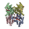
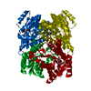
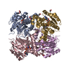
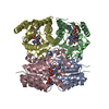
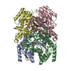
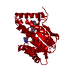
CRYSTAL STRUCTURE OF BETA-KETOACYL [ACYL CARRIER PROTEIN] REDUCTASE FROM E. COLI.
Components
BETA-KETOACYL [ACP] REDUCTASE
Keywords
OXIDOREDUCTASE / rossmann fold
Function / homology
Function and homology information
biotin biosynthetic process / 3-oxoacyl-[acyl-carrier-protein] reductase / 3-oxoacyl-[acyl-carrier-protein] reductase (NADPH) activity / fatty acid elongation / oxidoreductase activity, acting on the CH-OH group of donors, NAD or NADP as acceptor / lipid biosynthetic process / NAD binding / fatty acid biosynthetic process / NADP binding / metal ion binding ...biotin biosynthetic process / 3-oxoacyl-[acyl-carrier-protein] reductase / 3-oxoacyl-[acyl-carrier-protein] reductase (NADPH) activity / fatty acid elongation / oxidoreductase activity, acting on the CH-OH group of donors, NAD or NADP as acceptor / lipid biosynthetic process / NAD binding / fatty acid biosynthetic process / NADP binding / metal ion binding / identical protein binding / cytosol Similarity search - Function
HETEROGEN HOH 1-36 IS ASSOCIATED WITH CHAIN A. HOH 101-136 IS ASSOCIATED WITH CHAIN B. HOH 201-236 ...HETEROGEN HOH 1-36 IS ASSOCIATED WITH CHAIN A. HOH 101-136 IS ASSOCIATED WITH CHAIN B. HOH 201-236 IS ASSOCIATED WITH CHAIN C. HOH 301-336 IS ASSOCIATED WITH CHAIN D. HOH 401-436 IS ASSOCIATED WITH CHAIN E. HOH 501-536 IS ASSOCIATED WITH CHAIN F. HOH 601-636 IS ASSOCIATED WITH CHAIN G. HOH 701-736 IS ASSOCIATED WITH CHAIN H.
Mass: 25587.277 Da / Num. of mol.: 8 Source method: isolated from a genetically manipulated source Source: (gene. exp.) Escherichia coli (E. coli) / Production host: Escherichia coli (E. coli) References: UniProt: P0AEK2, 3-oxoacyl-[acyl-carrier-protein] reductase
In the structure databanks used in Yorodumi, some data are registered as the other names, "COVID-19 virus" and "2019-nCoV". Here are the details of the virus and the list of structure data.
Jan 31, 2019. EMDB accession codes are about to change! (news from PDBe EMDB page)
EMDB accession codes are about to change! (news from PDBe EMDB page)
The allocation of 4 digits for EMDB accession codes will soon come to an end. Whilst these codes will remain in use, new EMDB accession codes will include an additional digit and will expand incrementally as the available range of codes is exhausted. The current 4-digit format prefixed with “EMD-” (i.e. EMD-XXXX) will advance to a 5-digit format (i.e. EMD-XXXXX), and so on. It is currently estimated that the 4-digit codes will be depleted around Spring 2019, at which point the 5-digit format will come into force.
The EM Navigator/Yorodumi systems omit the EMD- prefix.
Related info.:Q: What is EMD? / ID/Accession-code notation in Yorodumi/EM Navigator
Yorodumi is a browser for structure data from EMDB, PDB, SASBDB, etc.
This page is also the successor to EM Navigator detail page, and also detail information page/front-end page for Omokage search.
The word "yorodu" (or yorozu) is an old Japanese word meaning "ten thousand". "mi" (miru) is to see.
Related info.:EMDB / PDB / SASBDB / Comparison of 3 databanks / Yorodumi Search / Aug 31, 2016. New EM Navigator & Yorodumi / Yorodumi Papers / Jmol/JSmol / Function and homology information / Changes in new EM Navigator and Yorodumi
 Movie
Movie Controller

 Yorodumi
 Open data
 Basic information
 Components
 Keywords
 Function and homology information
 X-RAY DIFFRACTION /
  Authors
 Citation
 Structure visualization
 Downloads & links
 Other downloads
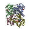


 PDBj
PDBj


 Assembly


 Mass: 18.015 Da / Num. of mol.: 349 / Source method: isolated from a natural source / Formula: H2O
 Sample preparation
 / Beamline: X25 / Wavelength: 0.97938, 0.95373
 Processing