 Movie
Movie Controller

[English] 日本語
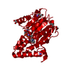
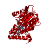
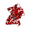
 Yorodumi
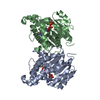
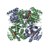
- PDB-1zid: LONG FATTY ACID CHAIN ENOYL-ACP REDUCTASE (INHA) IN COMPLEX WITH ... -
+ Open data

- Basic information
| Entry | Database: PDB / ID: 1zid | ||||||
|---|---|---|---|---|---|---|---|
| Title | LONG FATTY ACID CHAIN ENOYL-ACP REDUCTASE (INHA) IN COMPLEX WITH AN ISONICOTINIC-ACYL-NADH INHIBITOR | ||||||
 Components Components | ENOYL-[ACYL-CARRIER-PROTEIN] REDUCTASE | ||||||
 Keywords Keywords | OXIDOREDUCTASE / INHA ENZYME / ISONIAZID / MODIFIED NADH / ENOYL-ACP REDUCTASE / TUBERCULOSIS / MYCOLIC ACID BIOSYNTHESIS / Structural Genomics / PSI / Protein Structure Initiative / TB Structural Genomics Consortium / TBSGC | ||||||
| Function / homology |  Function and homology information Function and homology informationenoyl-[acyl-carrier-protein] reductase [NAD(P)H] activity / trans-2-enoyl-CoA reductase (NADH) activity / mycolic acid biosynthetic process / enoyl-[acyl-carrier-protein] reductase (NADH) / fatty acid elongation / enoyl-[acyl-carrier-protein] reductase (NADH) activity / NAD+ binding / peptidoglycan-based cell wall / fatty acid binding / fatty acid biosynthetic process ...enoyl-[acyl-carrier-protein] reductase [NAD(P)H] activity / trans-2-enoyl-CoA reductase (NADH) activity / mycolic acid biosynthetic process / enoyl-[acyl-carrier-protein] reductase (NADH) / fatty acid elongation / enoyl-[acyl-carrier-protein] reductase (NADH) activity / NAD+ binding / peptidoglycan-based cell wall / fatty acid binding / fatty acid biosynthetic process / response to antibiotic / plasma membrane Similarity search - Function | ||||||
| Biological species |   Mycobacterium tuberculosis (bacteria) Mycobacterium tuberculosis (bacteria) | ||||||
| Method |  X-RAY DIFFRACTION / INITIAL PHASES DERIVED FROM NATIVE STRUCTURE (PDB ENTRY 1ENY) / Resolution: 2.7 Å X-RAY DIFFRACTION / INITIAL PHASES DERIVED FROM NATIVE STRUCTURE (PDB ENTRY 1ENY) / Resolution: 2.7 Å | ||||||
 Authors Authors | Rozwarski, D.A. / Jacobs Jr., W.R. / Sacchettini, J.C. / TB Structural Genomics Consortium (TBSGC) | ||||||
 Citation Citation | Journal: Science / Year: 1998 Title: Modification of the NADH of the isoniazid target (InhA) from Mycobacterium tuberculosis. Authors: Rozwarski, D.A. / Grant, G.A. / Barton, D.H. / Jacobs Jr., W.R. / Sacchettini, J.C. #1: Journal: Science / Year: 1995Title: Crystal Structure and Function of the Isoniazid Target of Mycobacterium Tuberculosis Authors: Dessen, A. / Quemard, A. / Blanchard, J.S. / Jacobs Junior, W.R. / Sacchettini, J.C. | ||||||
| History |
|
- Structure visualization
| Structure viewer | Molecule: MolmilJmol/JSmol |
|---|
- Downloads & links
-Download
| PDBx/mmCIF format | 1zid.cif.gz | 65.1 KB | Display | PDBx/mmCIF format |
|---|---|---|---|---|
| PDB format | pdb1zid.ent.gz | 47.7 KB | Display | PDB format |
| PDBx/mmJSON format | 1zid.json.gz | Tree view | PDBx/mmJSON format | |
| Others |  Other downloads Other downloads |
-Validation report
| Arichive directory | https://data.pdbj.org/pub/pdb/validation_reports/zi/1zidftp://data.pdbj.org/pub/pdb/validation_reports/zi/1zid | HTTPS FTP |
|---|
-Related structure data
| Related structure data | 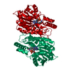 1enyS S: Starting model for refinement |
|---|---|
| Similar structure data | |
| Other databases |

-Links
 PDBj
PDBj

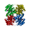
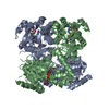
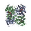
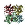
- Assembly
| Deposited unit | 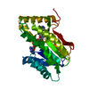
| ||||||||
|---|---|---|---|---|---|---|---|---|---|
| 1 | 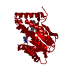
| ||||||||
| Unit cell |
|
-Components
| #1: Protein | Mass: 28393.562 Da / Num. of mol.: 1 / Mutation: T2A Source method: isolated from a genetically manipulated source Source: (gene. exp.) Mycobacterium tuberculosis (bacteria) / Species (production host): Escherichia coli / Production host: References: UniProt: P0A5Y6, UniProt: P9WGR1*PLUS, enoyl-[acyl-carrier-protein] reductase (NADH) |
|---|---|
| #2: Chemical | ChemComp-ZID /   Mass: 768.519 Da / Num. of mol.: 1 / Source method: obtained synthetically / Formula: C27H30N8O15P2 Mass: 768.519 Da / Num. of mol.: 1 / Source method: obtained synthetically / Formula: C27H30N8O15P2 |
| #3: Water | ChemComp-HOH /  Mass: 18.015 Da / Num. of mol.: 68 / Source method: isolated from a natural source / Formula: H2O Mass: 18.015 Da / Num. of mol.: 68 / Source method: isolated from a natural source / Formula: H2O |
-Experimental details
-Experiment
| Experiment | Method: X-RAY DIFFRACTION / Number of used crystals: 1 |
|---|
- Sample preparation
| Crystal | Density Matthews: 3.58 Å3/Da / Density % sol: 65.6 % | ||||||||||||||||||||||||||||||
|---|---|---|---|---|---|---|---|---|---|---|---|---|---|---|---|---|---|---|---|---|---|---|---|---|---|---|---|---|---|---|---|
| Crystal grow | pH: 7.5 Details: 12% MPD, 4% DMSO, 50MM NACITRATE, 100 MM HEPES, PH 7.5 | ||||||||||||||||||||||||||||||
| Crystal grow | *PLUS Method: vapor diffusion, hanging drop | ||||||||||||||||||||||||||||||
| Components of the solutions | *PLUS
|
-Data collection
| Diffraction | Mean temperature: 298 K |
|---|---|
| Diffraction source | Source: ROTATING ANODE / Type: RIGAKU / Wavelength: 1.5418 |
| Detector | Type: MACSCIENCE / Detector: IMAGE PLATE / Date: Nov 1, 1996 / Details: MIRRORS |
| Radiation | Monochromator: 0.005 MM NI FILTER / Monochromatic (M) / Laue (L): M / Scattering type: x-ray |
| Radiation wavelength | Wavelength: 1.5418 Å / Relative weight: 1 |
| Reflection | Resolution: 2.7→10 Å / Num. obs: 10621 / % possible obs: 90.7 % / Observed criterion σ(I): 1 / Rsym value: 0.164 / Net I/σ(I): 11.3 |
| Reflection shell | Resolution: 2.7→2.77 Å / Mean I/σ(I) obs: 2.4 / Rsym value: 0.343 / % possible all: 49.5 |
| Reflection | *PLUS Rmerge(I) obs: 0.164 |
| Reflection shell | *PLUS % possible obs: 50 % / Rmerge(I) obs: 0.343 |
- Processing
| Software |
| ||||||||||||||||||||||||||||||||||||||||||||||||||||||||||||||||||||||||||||||||
|---|---|---|---|---|---|---|---|---|---|---|---|---|---|---|---|---|---|---|---|---|---|---|---|---|---|---|---|---|---|---|---|---|---|---|---|---|---|---|---|---|---|---|---|---|---|---|---|---|---|---|---|---|---|---|---|---|---|---|---|---|---|---|---|---|---|---|---|---|---|---|---|---|---|---|---|---|---|---|---|---|---|
| Refinement | Method to determine structure: INITIAL PHASES DERIVED FROM NATIVE STRUCTURE (PDB ENTRY 1ENY) Starting model: NATIVE STRUCTURE (PDB ENTRY 1ENY) Resolution: 2.7→10 Å / Data cutoff high absF: 100000 / Data cutoff low absF: 0.1 / Isotropic thermal model: RESTRAINED / Cross valid method: THROUGHOUT / σ(F): 2
| ||||||||||||||||||||||||||||||||||||||||||||||||||||||||||||||||||||||||||||||||
| Displacement parameters | Biso mean: 24.3 Å2 | ||||||||||||||||||||||||||||||||||||||||||||||||||||||||||||||||||||||||||||||||
| Refine analyze |
| ||||||||||||||||||||||||||||||||||||||||||||||||||||||||||||||||||||||||||||||||
| Refinement step | Cycle: LAST / Resolution: 2.7→10 Å
| ||||||||||||||||||||||||||||||||||||||||||||||||||||||||||||||||||||||||||||||||
| Refine LS restraints |
| ||||||||||||||||||||||||||||||||||||||||||||||||||||||||||||||||||||||||||||||||
| LS refinement shell | Resolution: 2.7→2.77 Å / Total num. of bins used: 12
| ||||||||||||||||||||||||||||||||||||||||||||||||||||||||||||||||||||||||||||||||
| Xplor file |
| ||||||||||||||||||||||||||||||||||||||||||||||||||||||||||||||||||||||||||||||||
| Software | *PLUS Name: X-PLOR / Version: 3.1 / Classification: refinement | ||||||||||||||||||||||||||||||||||||||||||||||||||||||||||||||||||||||||||||||||
| Refine LS restraints | *PLUS
|
