 Movie
Movie Controller

[English] 日本語
 Yorodumi
- PDB-3wye: Crystal Structure of chimeric engineered (2S,3S)-butanediol dehyd... -
+ Open data

- Basic information
| Entry | Database: PDB / ID: 3wye | ||||||
|---|---|---|---|---|---|---|---|
| Title | Crystal Structure of chimeric engineered (2S,3S)-butanediol dehydrogenase complexed with NAD+ | ||||||
 Components Components | Diacetyl reductase [(S)-acetoin forming],L-2,3-butanediol dehydrogenase,Diacetyl reductase [(S)-acetoin forming],L-2,3-butanediol dehydrogenase,Diacetyl reductase [(S)-acetoin forming],L-2,3-butanediol dehydrogenase,Diacetyl reductase [(S)-acetoin forming] | ||||||
 Keywords Keywords | OXIDOREDUCTASE / butanediol dehydrogenase / domain chimera / NAD+ complex / short-chain dehydrogenase/reductase family / Rossmann Fold / NAD+ Binding / Oxidation/reduction | ||||||
| Function / homology |  Function and homology information Function and homology information(S,S)-butanediol dehydrogenase / (S,S)-butanediol dehydrogenase activity / acetoin metabolic process / diacetyl reductase [(S)-acetoin forming] / butanediol metabolic process / diacetyl reductase ((S)-acetoin forming) (NAD+) activity / acetoin catabolic process / NADH binding / NAD+ binding / quinone binding ...(S,S)-butanediol dehydrogenase / (S,S)-butanediol dehydrogenase activity / acetoin metabolic process / diacetyl reductase [(S)-acetoin forming] / butanediol metabolic process / diacetyl reductase ((S)-acetoin forming) (NAD+) activity / acetoin catabolic process / NADH binding / NAD+ binding / quinone binding / fatty acid biosynthetic process / protein homotetramerization Similarity search - Function | ||||||
| Biological species |  Klebsiella pneumoniae (bacteria)Corynebacterium glutamicum (bacteria) Klebsiella pneumoniae (bacteria)Corynebacterium glutamicum (bacteria) | ||||||
| Method |  X-RAY DIFFRACTION / SYNCHROTRON / MOLECULAR REPLACEMENT / Resolution: 1.58 Å X-RAY DIFFRACTION / SYNCHROTRON / MOLECULAR REPLACEMENT / Resolution: 1.58 Å | ||||||
 Authors Authors | Shimegi, T. / Oyama, T. / Kusunoki, M. / Ui, S. | ||||||
 Citation Citation | Journal: To be Published Title: Crystal Structure of chimeric engineered (2S,3S)-butanediol dehydrogenase complexed with NAD+ Authors: Shimegi, T. / Oyama, T. / Kusunoki, M. / Ui, S. | ||||||
| History |
|
- Structure visualization
| Structure viewer | Molecule: MolmilJmol/JSmol |
|---|
- Downloads & links
-Download
| PDBx/mmCIF format | 3wye.cif.gz | 107 KB | Display | PDBx/mmCIF format |
|---|---|---|---|---|
| PDB format | pdb3wye.ent.gz | 82.3 KB | Display | PDB format |
| PDBx/mmJSON format | 3wye.json.gz | Tree view | PDBx/mmJSON format | |
| Others |  Other downloads Other downloads |
-Validation report
| Arichive directory | https://data.pdbj.org/pub/pdb/validation_reports/wy/3wyeftp://data.pdbj.org/pub/pdb/validation_reports/wy/3wye | HTTPS FTP |
|---|
-Related structure data
| Related structure data | |
|---|---|
| Similar structure data |


-Links
 PDBj
PDBj


- Assembly
| Deposited unit | 
| ||||||||
|---|---|---|---|---|---|---|---|---|---|
| 1 | 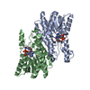
| ||||||||
| Unit cell |
| ||||||||
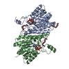
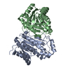
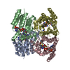
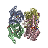
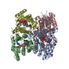
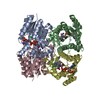
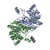
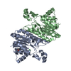
| Details | The biological assembly is a tetramer generated from the dimer in the asymmetric unit by two fold symmetry operation: x, -y, -z. |
-Components
| #1: Protein | Mass: 26720.445 Da / Num. of mol.: 2 Mutation: R52H, M54V,R52H, M54V,R52H, M54V,R52H, M54V,R52H, M54V,R52H, M54V,R52H, M54V Source method: isolated from a genetically manipulated source Details: Residues 84-118, 135-161 and 182-236 derived from Corynebacterium glutamicum (2S,3S)-butanediol dehydrogenase and the remaining resides from Klebsiella pneumoniae (2R,3S)-butanediol dehydrogenase Source: (gene. exp.) Klebsiella pneumoniae (bacteria), (gene. exp.) Corynebacterium glutamicum (bacteria)Gene: budC / Plasmid: pET11a / Production host: References: UniProt: Q48436, UniProt: Q9ZNN8, diacetyl reductase [(S)-acetoin forming], (S,S)-butanediol dehydrogenase #2: Chemical |   Mass: 663.425 Da / Num. of mol.: 2 / Source method: obtained synthetically / Formula: C21H27N7O14P2 / Comment: NAD*YM Mass: 663.425 Da / Num. of mol.: 2 / Source method: obtained synthetically / Formula: C21H27N7O14P2 / Comment: NAD*YM#3: Water | ChemComp-HOH / |  Mass: 18.015 Da / Num. of mol.: 140 / Source method: isolated from a natural source / Formula: H2O Mass: 18.015 Da / Num. of mol.: 140 / Source method: isolated from a natural source / Formula: H2O |
|---|
-Experimental details
-Experiment
| Experiment | Method: X-RAY DIFFRACTION / Number of used crystals: 1 |
|---|
- Sample preparation
| Crystal | Density Matthews: 2.25 Å3/Da / Density % sol: 45.23 % / Mosaicity: 0.532 ° |
|---|---|
| Crystal grow | Temperature: 293 K / Method: vapor diffusion, hanging drop / pH: 6.4 Details: 17% PEG6000, 100mM MES, 15% glycerol, 1% 2-mercaptoethanol, pH 6.4, vapor diffusion, hanging drop, temperature 293K |
-Data collection
| Diffraction | Mean temperature: 95 K | ||||||||||||||||||||||||||||||||||||||||||||||||||||||||||||||||||||||||||||||||||||||||||||||||||||||||||||||||||||||||||||||
|---|---|---|---|---|---|---|---|---|---|---|---|---|---|---|---|---|---|---|---|---|---|---|---|---|---|---|---|---|---|---|---|---|---|---|---|---|---|---|---|---|---|---|---|---|---|---|---|---|---|---|---|---|---|---|---|---|---|---|---|---|---|---|---|---|---|---|---|---|---|---|---|---|---|---|---|---|---|---|---|---|---|---|---|---|---|---|---|---|---|---|---|---|---|---|---|---|---|---|---|---|---|---|---|---|---|---|---|---|---|---|---|---|---|---|---|---|---|---|---|---|---|---|---|---|---|---|---|
| Diffraction source | Source: SYNCHROTRON / Site: Photon Factory  / Beamline: AR-NE3A / Wavelength: 1 Å / Beamline: AR-NE3A / Wavelength: 1 Å | ||||||||||||||||||||||||||||||||||||||||||||||||||||||||||||||||||||||||||||||||||||||||||||||||||||||||||||||||||||||||||||||
| Detector | Type: ADSC QUANTUM 210r / Detector: CCD / Date: May 26, 2013 | ||||||||||||||||||||||||||||||||||||||||||||||||||||||||||||||||||||||||||||||||||||||||||||||||||||||||||||||||||||||||||||||
| Radiation | Monochromator: Si 111 CHANNEL / Protocol: SINGLE WAVELENGTH / Monochromatic (M) / Laue (L): M / Scattering type: x-ray | ||||||||||||||||||||||||||||||||||||||||||||||||||||||||||||||||||||||||||||||||||||||||||||||||||||||||||||||||||||||||||||||
| Radiation wavelength | Wavelength: 1 Å / Relative weight: 1 | ||||||||||||||||||||||||||||||||||||||||||||||||||||||||||||||||||||||||||||||||||||||||||||||||||||||||||||||||||||||||||||||
| Reflection | Resolution: 1.578→50 Å / Num. obs: 65845 / % possible obs: 99.4 % / Redundancy: 8.9 % / Biso Wilson estimate: 18.88 Å2 / Rmerge(I) obs: 0.051 / Χ2: 1.027 / Net I/σ(I): 19.2 | ||||||||||||||||||||||||||||||||||||||||||||||||||||||||||||||||||||||||||||||||||||||||||||||||||||||||||||||||||||||||||||||
| Reflection shell | Diffraction-ID: 1 / Rejects: _
|
- Processing
| Software |
| |||||||||||||||||||||||||||||||||||||||||||||||||||||||||||||||||||||||||||||||||||||||||||||||||||||||||||||||||||||||||||||||||||||||||||||||||||||||||||||||||||||||||||||||
|---|---|---|---|---|---|---|---|---|---|---|---|---|---|---|---|---|---|---|---|---|---|---|---|---|---|---|---|---|---|---|---|---|---|---|---|---|---|---|---|---|---|---|---|---|---|---|---|---|---|---|---|---|---|---|---|---|---|---|---|---|---|---|---|---|---|---|---|---|---|---|---|---|---|---|---|---|---|---|---|---|---|---|---|---|---|---|---|---|---|---|---|---|---|---|---|---|---|---|---|---|---|---|---|---|---|---|---|---|---|---|---|---|---|---|---|---|---|---|---|---|---|---|---|---|---|---|---|---|---|---|---|---|---|---|---|---|---|---|---|---|---|---|---|---|---|---|---|---|---|---|---|---|---|---|---|---|---|---|---|---|---|---|---|---|---|---|---|---|---|---|---|---|---|---|---|---|
| Refinement | Method to determine structure: MOLECULAR REPLACEMENT / Resolution: 1.58→39.279 Å / SU ML: 0.13 / Isotropic thermal model: Isotropic / σ(F): 1.35 / Phase error: 20.18 / Stereochemistry target values: ML
| |||||||||||||||||||||||||||||||||||||||||||||||||||||||||||||||||||||||||||||||||||||||||||||||||||||||||||||||||||||||||||||||||||||||||||||||||||||||||||||||||||||||||||||||
| Solvent computation | Shrinkage radii: 0.9 Å / VDW probe radii: 1.11 Å / Solvent model: FLAT BULK SOLVENT MODEL | |||||||||||||||||||||||||||||||||||||||||||||||||||||||||||||||||||||||||||||||||||||||||||||||||||||||||||||||||||||||||||||||||||||||||||||||||||||||||||||||||||||||||||||||
| Displacement parameters | Biso max: 113.04 Å2 / Biso mean: 25.5213 Å2 / Biso min: 13.09 Å2 | |||||||||||||||||||||||||||||||||||||||||||||||||||||||||||||||||||||||||||||||||||||||||||||||||||||||||||||||||||||||||||||||||||||||||||||||||||||||||||||||||||||||||||||||
| Refinement step | Cycle: LAST / Resolution: 1.58→39.279 Å
| |||||||||||||||||||||||||||||||||||||||||||||||||||||||||||||||||||||||||||||||||||||||||||||||||||||||||||||||||||||||||||||||||||||||||||||||||||||||||||||||||||||||||||||||
| Refine LS restraints |
| |||||||||||||||||||||||||||||||||||||||||||||||||||||||||||||||||||||||||||||||||||||||||||||||||||||||||||||||||||||||||||||||||||||||||||||||||||||||||||||||||||||||||||||||
| LS refinement shell | Refine-ID: X-RAY DIFFRACTION / Total num. of bins used: 24
|
