 Movie
Movie Controller

[English] 日本語
 Yorodumi
- PDB-6uds: Crystal structure of a putative 3-oxoacyl-ACP reductase (FabG) fr... -
+ Open data

- Basic information
| Entry | Database: PDB / ID: 6uds | ||||||
|---|---|---|---|---|---|---|---|
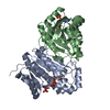
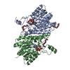
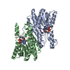
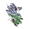
| Title | Crystal structure of a putative 3-oxoacyl-ACP reductase (FabG) from Acinetobacter baumannii | ||||||
 Components Components | 3-oxoacyl-(Acyl-carrier-protein) reductase | ||||||
 Keywords Keywords | OXIDOREDUCTASE / FabG / SDR / reductase / Rossmann Fold | ||||||
| Function / homology |  Function and homology information Function and homology information3-oxoacyl-[acyl-carrier-protein] reductase / 3-oxoacyl-[acyl-carrier-protein] reductase (NADPH) activity / fatty acid elongation / NAD binding Similarity search - Function | ||||||
| Biological species |  Acinetobacter baumannii (bacteria) Acinetobacter baumannii (bacteria) | ||||||
| Method |  X-RAY DIFFRACTION / SYNCHROTRON / MOLECULAR REPLACEMENT / Resolution: 1.9 Å X-RAY DIFFRACTION / SYNCHROTRON / MOLECULAR REPLACEMENT / Resolution: 1.9 Å | ||||||
 Authors Authors | Forwood, J.K. / Cross, E.M. | ||||||
 Citation Citation | Journal: Sci Rep / Year: 2021 Title: Insights into Acinetobacter baumannii fatty acid synthesis 3-oxoacyl-ACP reductases. Authors: Cross, E.M. / Adams, F.G. / Waters, J.K. / Aragao, D. / Eijkelkamp, B.A. / Forwood, J.K. | ||||||
| History |
|
- Structure visualization
| Structure viewer | Molecule: MolmilJmol/JSmol |
|---|
- Downloads & links
-Download
| PDBx/mmCIF format | 6uds.cif.gz | 226 KB | Display | PDBx/mmCIF format |
|---|---|---|---|---|
| PDB format | pdb6uds.ent.gz | 146.3 KB | Display | PDB format |
| PDBx/mmJSON format | 6uds.json.gz | Tree view | PDBx/mmJSON format | |
| Others |  Other downloads Other downloads |
-Validation report
| Arichive directory | https://data.pdbj.org/pub/pdb/validation_reports/ud/6udsftp://data.pdbj.org/pub/pdb/validation_reports/ud/6uds | HTTPS FTP |
|---|
-Related structure data
| Related structure data |  6nrpC  6uutC  6uuvC  6wprC  4wjzS S: Starting model for refinement C: citing same article ( |
|---|---|
| Similar structure data |

-Links
 PDBj
PDBj


- Assembly
| Deposited unit | 
| |||||||||||||||||||||||||||||||||||||||||||||||||||||||||||||
|---|---|---|---|---|---|---|---|---|---|---|---|---|---|---|---|---|---|---|---|---|---|---|---|---|---|---|---|---|---|---|---|---|---|---|---|---|---|---|---|---|---|---|---|---|---|---|---|---|---|---|---|---|---|---|---|---|---|---|---|---|---|---|
| 1 | 
| |||||||||||||||||||||||||||||||||||||||||||||||||||||||||||||
| Unit cell |
| |||||||||||||||||||||||||||||||||||||||||||||||||||||||||||||
| Components on special symmetry positions |
| |||||||||||||||||||||||||||||||||||||||||||||||||||||||||||||
| Noncrystallographic symmetry (NCS) | NCS domain:
NCS domain segments: Ens-ID: 1
|
-Components
| #1: Protein | Mass: 28870.582 Da / Num. of mol.: 2 Source method: isolated from a genetically manipulated source Source: (gene. exp.) Acinetobacter baumannii (bacteria)Gene: fabG_9, fabG, fabG_10, fabG_11, fabG_12, fabG_2, fabG_3, fabG_5, fabG_7, fabG_8, A7M79_02495, A7M90_13795, A7N09_01015, AB719_00430, ABUW_3108, B4R90_07750, B9X91_07565, B9X95_05710, BGC29_ ...Gene: fabG_9, fabG, fabG_10, fabG_11, fabG_12, fabG_2, fabG_3, fabG_5, fabG_7, fabG_8, A7M79_02495, A7M90_13795, A7N09_01015, AB719_00430, ABUW_3108, B4R90_07750, B9X91_07565, B9X95_05710, BGC29_09155, BWP00_12180, C2U32_18365, C3415_14660, CBE85_08910, CBI29_00841, CEJ63_15165, CHQ89_11440, CPI82_11015, CSB70_0489, CYQ93_20585, CYQ93_22950, D3X57_12365, DCD77_08675, DGS69_00745, DOL94_00645, DVA79_16530, DWA21_01645, E2535_15965, E4664_12920, E5D09_05595, EA682_12370, EA685_06995, EGM95_16765, EHF38_14445, EJB02_04655, EKS29_20225, EWO92_12305, EWO96_16390, EWP49_14850, FDF20_02980, LV38_02926, NCTC13305_01645, NT90_07375, SAMEA104305229_02428, SAMEA104305242_00720, SAMEA104305268_02089, SAMEA104305283_02395, SAMEA104305292_02313, SAMEA104305318_02347, SAMEA104305320_01823, SAMEA104305325_02271, SAMEA104305337_03003, SAMEA104305351_01934 Production host: References: UniProt: V5VHN7, 3-oxoacyl-[acyl-carrier-protein] reductase #2: Water | ChemComp-HOH / |  Mass: 18.015 Da / Num. of mol.: 326 / Source method: isolated from a natural source / Formula: H2O Mass: 18.015 Da / Num. of mol.: 326 / Source method: isolated from a natural source / Formula: H2O |
|---|
-Experimental details
-Experiment
| Experiment | Method: X-RAY DIFFRACTION / Number of used crystals: 1 |
|---|
- Sample preparation
| Crystal | Density Matthews: 2.5 Å3/Da / Density % sol: 50.8 % |
|---|---|
| Crystal grow | Temperature: 296.15 K / Method: vapor diffusion, hanging drop / Details: 0.1 M Tris, pH 8.0, 2 M ammonium sulfate |
-Data collection
| Diffraction | Mean temperature: 100 K / Serial crystal experiment: N |
|---|---|
| Diffraction source | Source: SYNCHROTRON / Site: Australian Synchrotron  / Beamline: MX2 / Wavelength: 0.95366 Å / Beamline: MX2 / Wavelength: 0.95366 Å |
| Detector | Type: DECTRIS EIGER X 16M / Detector: PIXEL / Date: Feb 7, 2018 |
| Radiation | Protocol: SINGLE WAVELENGTH / Monochromatic (M) / Laue (L): M / Scattering type: x-ray |
| Radiation wavelength | Wavelength: 0.95366 Å / Relative weight: 1 |
| Reflection | Resolution: 1.9→29.13 Å / Num. obs: 46833 / % possible obs: 99.8 % / Redundancy: 18 % / Biso Wilson estimate: 32.53 Å2 / CC1/2: 0.997 / Rmerge(I) obs: 0.164 / Rpim(I) all: 0.04 / Rrim(I) all: 0.169 / Net I/σ(I): 9.6 |
| Reflection shell | Resolution: 1.9→1.94 Å / Rmerge(I) obs: 1.128 / Mean I/σ(I) obs: 2 / Num. unique obs: 3036 / CC1/2: 0.781 / Rpim(I) all: 0.288 / Rrim(I) all: 1.166 |
- Processing
| Software |
| |||||||||||||||||||||||||||||||||||||||||||||||||||||||||||||||||||||||||||||||||||||||||||||||||||||||||||||||||||||||||||||||||||||
|---|---|---|---|---|---|---|---|---|---|---|---|---|---|---|---|---|---|---|---|---|---|---|---|---|---|---|---|---|---|---|---|---|---|---|---|---|---|---|---|---|---|---|---|---|---|---|---|---|---|---|---|---|---|---|---|---|---|---|---|---|---|---|---|---|---|---|---|---|---|---|---|---|---|---|---|---|---|---|---|---|---|---|---|---|---|---|---|---|---|---|---|---|---|---|---|---|---|---|---|---|---|---|---|---|---|---|---|---|---|---|---|---|---|---|---|---|---|---|---|---|---|---|---|---|---|---|---|---|---|---|---|---|---|---|
| Refinement | Method to determine structure: MOLECULAR REPLACEMENT Starting model: PDB entry 4WJZ Resolution: 1.9→29.13 Å / SU ML: 0.1957 / Cross valid method: FREE R-VALUE / σ(F): 1.34 / Phase error: 21.9984
| |||||||||||||||||||||||||||||||||||||||||||||||||||||||||||||||||||||||||||||||||||||||||||||||||||||||||||||||||||||||||||||||||||||
| Solvent computation | Shrinkage radii: 0.9 Å / VDW probe radii: 1.11 Å | |||||||||||||||||||||||||||||||||||||||||||||||||||||||||||||||||||||||||||||||||||||||||||||||||||||||||||||||||||||||||||||||||||||
| Displacement parameters | Biso mean: 39.07 Å2 | |||||||||||||||||||||||||||||||||||||||||||||||||||||||||||||||||||||||||||||||||||||||||||||||||||||||||||||||||||||||||||||||||||||
| Refinement step | Cycle: LAST / Resolution: 1.9→29.13 Å
| |||||||||||||||||||||||||||||||||||||||||||||||||||||||||||||||||||||||||||||||||||||||||||||||||||||||||||||||||||||||||||||||||||||
| Refine LS restraints |
| |||||||||||||||||||||||||||||||||||||||||||||||||||||||||||||||||||||||||||||||||||||||||||||||||||||||||||||||||||||||||||||||||||||
| LS refinement shell |
|
