 Movie
Movie Controller

[English] 日本語
 Yorodumi
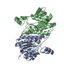
- PDB-6wpr: Crystal structure of a putative 3-oxoacyl-ACP reductase (FabG) wi... -
+ Open data

- Basic information
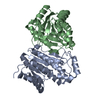
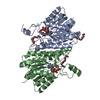
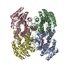
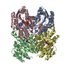
| Entry | Database: PDB / ID: 6wpr | ||||||
|---|---|---|---|---|---|---|---|
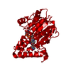
| Title | Crystal structure of a putative 3-oxoacyl-ACP reductase (FabG) with NADP(H) from Acinetobacter baumannii | ||||||
 Components Components | 3-oxoacyl-[acyl-carrier-protein] reductase | ||||||
 Keywords Keywords | OXIDOREDUCTASE / FabG / reductase / SDR / Rossmann Fold | ||||||
| Function / homology |  Function and homology information Function and homology information3-oxoacyl-[acyl-carrier-protein] reductase / 3-oxoacyl-[acyl-carrier-protein] reductase (NADPH) activity / fatty acid elongation / NAD binding Similarity search - Function | ||||||
| Biological species |  Acinetobacter baumannii (bacteria) Acinetobacter baumannii (bacteria) | ||||||
| Method |  X-RAY DIFFRACTION / SYNCHROTRON / MOLECULAR REPLACEMENT / Resolution: 1.85 Å X-RAY DIFFRACTION / SYNCHROTRON / MOLECULAR REPLACEMENT / Resolution: 1.85 Å | ||||||
 Authors Authors | Cross, E.M. / Forwood, J.K. | ||||||
 Citation Citation | Journal: Sci Rep / Year: 2021 Title: Insights into Acinetobacter baumannii fatty acid synthesis 3-oxoacyl-ACP reductases. Authors: Cross, E.M. / Adams, F.G. / Waters, J.K. / Aragao, D. / Eijkelkamp, B.A. / Forwood, J.K. | ||||||
| History |
|
- Structure visualization
| Structure viewer | Molecule: MolmilJmol/JSmol |
|---|
- Downloads & links
-Download
| PDBx/mmCIF format | 6wpr.cif.gz | 225.4 KB | Display | PDBx/mmCIF format |
|---|---|---|---|---|
| PDB format | pdb6wpr.ent.gz | 156.1 KB | Display | PDB format |
| PDBx/mmJSON format | 6wpr.json.gz | Tree view | PDBx/mmJSON format | |
| Others |  Other downloads Other downloads |
-Validation report
| Arichive directory | https://data.pdbj.org/pub/pdb/validation_reports/wp/6wprftp://data.pdbj.org/pub/pdb/validation_reports/wp/6wpr | HTTPS FTP |
|---|
-Related structure data
| Related structure data |  6nrpC  6udsSC 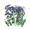 6uutC  6uuvC S: Starting model for refinement C: citing same article ( |
|---|---|
| Similar structure data |


-Links
 PDBj
PDBj


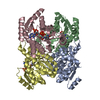
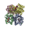
- Assembly
| Deposited unit | 
| ||||||||||||
|---|---|---|---|---|---|---|---|---|---|---|---|---|---|
| 1 | 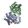
| ||||||||||||
| Unit cell |
| ||||||||||||
| Components on special symmetry positions |
|
-Components
| #1: Protein | Mass: 26126.660 Da / Num. of mol.: 2 Source method: isolated from a genetically manipulated source Source: (gene. exp.) Acinetobacter baumannii (bacteria)Gene: fabG_9, fabG, fabG_5, fabG_8, A7M79_02495, A7M90_13795, A9843_07395, AA954_11685, ABCAM1_0760, ABUW_3108, APD33_00335, B4R90_07750, B9W69_04055, B9X95_05710, BGC29_09155, C2U32_18365, CEJ64_ ...Gene: fabG_9, fabG, fabG_5, fabG_8, A7M79_02495, A7M90_13795, A9843_07395, AA954_11685, ABCAM1_0760, ABUW_3108, APD33_00335, B4R90_07750, B9W69_04055, B9X95_05710, BGC29_09155, C2U32_18365, CEJ64_13010, CHQ89_11440, CPI82_11015, CSB70_0489, DLI69_06485, DLI75_02490, DOL94_00645, DVA79_16530, E2533_13485, E2536_16310, E5294_15795, E5979_01830, EA685_06995, EA746_003475, EKS29_20225, EWO92_12305, EWO96_16390, EWP49_14850, FD887_09475, FD913_04775, FJU36_15155, FJU42_04975, FJU76_14990, FJU79_09000, FJU87_10860, FJV14_09530, LV38_02926, NCTC13305_01645, NCTC13420_02176, NT90_07375, SAMEA104305318_02347, SAMEA104305351_01934 Production host: References: UniProt: V5VHN7, 3-oxoacyl-[acyl-carrier-protein] reductase #2: Chemical |   Mass: 745.421 Da / Num. of mol.: 2 / Source method: obtained synthetically / Formula: C21H30N7O17P3 / Feature type: SUBJECT OF INVESTIGATION Mass: 745.421 Da / Num. of mol.: 2 / Source method: obtained synthetically / Formula: C21H30N7O17P3 / Feature type: SUBJECT OF INVESTIGATION#3: Water | ChemComp-HOH / |  Mass: 18.015 Da / Num. of mol.: 495 / Source method: isolated from a natural source / Formula: H2O Mass: 18.015 Da / Num. of mol.: 495 / Source method: isolated from a natural source / Formula: H2OHas ligand of interest | Y | |
|---|
-Experimental details
-Experiment
| Experiment | Method: X-RAY DIFFRACTION / Number of used crystals: 1 |
|---|
- Sample preparation
| Crystal | Density Matthews: 2.77 Å3/Da / Density % sol: 55.64 % |
|---|---|
| Crystal grow | Temperature: 296.15 K / Method: vapor diffusion, hanging drop / Details: 0.1 M Tris, pH 8.0, 2 M ammonium sulfate |
-Data collection
| Diffraction | Mean temperature: 100 K / Serial crystal experiment: N |
|---|---|
| Diffraction source | Source: SYNCHROTRON / Site: Australian Synchrotron  / Beamline: MX1 / Wavelength: 0.9537 Å / Beamline: MX1 / Wavelength: 0.9537 Å |
| Detector | Type: ADSC QUANTUM 210r / Detector: CCD / Date: Jul 28, 2018 |
| Radiation | Monochromator: double crystal Si(111) / Protocol: SINGLE WAVELENGTH / Monochromatic (M) / Laue (L): M / Scattering type: x-ray |
| Radiation wavelength | Wavelength: 0.9537 Å / Relative weight: 1 |
| Reflection | Resolution: 1.85→50.55 Å / Num. obs: 51013 / % possible obs: 100 % / Redundancy: 14.3 % / Biso Wilson estimate: 18.94 Å2 / CC1/2: 1 / Rmerge(I) obs: 0.077 / Rpim(I) all: 0.021 / Rrim(I) all: 0.08 / Net I/σ(I): 26.3 |
| Reflection shell | Resolution: 1.85→1.89 Å / Rmerge(I) obs: 0.584 / Mean I/σ(I) obs: 4.9 / Num. unique obs: 3085 / CC1/2: 0.939 / Rpim(I) all: 0.158 / Rrim(I) all: 0.606 |
- Processing
| Software |
| |||||||||||||||||||||||||||||||||||||||||||||||||||||||||||||||||||||||||||||||||||||||||||||||||||||||||||||||||||||||||||||||||||||
|---|---|---|---|---|---|---|---|---|---|---|---|---|---|---|---|---|---|---|---|---|---|---|---|---|---|---|---|---|---|---|---|---|---|---|---|---|---|---|---|---|---|---|---|---|---|---|---|---|---|---|---|---|---|---|---|---|---|---|---|---|---|---|---|---|---|---|---|---|---|---|---|---|---|---|---|---|---|---|---|---|---|---|---|---|---|---|---|---|---|---|---|---|---|---|---|---|---|---|---|---|---|---|---|---|---|---|---|---|---|---|---|---|---|---|---|---|---|---|---|---|---|---|---|---|---|---|---|---|---|---|---|---|---|---|
| Refinement | Method to determine structure: MOLECULAR REPLACEMENT Starting model: PDB entry 6UDS Resolution: 1.85→47.91 Å / SU ML: 0.1834 / Cross valid method: FREE R-VALUE / σ(F): 1.34 / Phase error: 16.9494 Stereochemistry target values: GeoStd + Monomer Library + CDL v1.2
| |||||||||||||||||||||||||||||||||||||||||||||||||||||||||||||||||||||||||||||||||||||||||||||||||||||||||||||||||||||||||||||||||||||
| Solvent computation | Shrinkage radii: 0.9 Å / VDW probe radii: 1.11 Å / Solvent model: FLAT BULK SOLVENT MODEL | |||||||||||||||||||||||||||||||||||||||||||||||||||||||||||||||||||||||||||||||||||||||||||||||||||||||||||||||||||||||||||||||||||||
| Displacement parameters | Biso mean: 22.53 Å2 | |||||||||||||||||||||||||||||||||||||||||||||||||||||||||||||||||||||||||||||||||||||||||||||||||||||||||||||||||||||||||||||||||||||
| Refinement step | Cycle: LAST / Resolution: 1.85→47.91 Å
| |||||||||||||||||||||||||||||||||||||||||||||||||||||||||||||||||||||||||||||||||||||||||||||||||||||||||||||||||||||||||||||||||||||
| Refine LS restraints |
| |||||||||||||||||||||||||||||||||||||||||||||||||||||||||||||||||||||||||||||||||||||||||||||||||||||||||||||||||||||||||||||||||||||
| LS refinement shell |
|
