 Movie
Movie Controller

[English] 日本語
 Yorodumi
- PDB-3grk: Crystal structure of short chain dehydrogenase reductase SDR gluc... -
+ Open data

- Basic information
| Entry | Database: PDB / ID: 3grk | ||||||
|---|---|---|---|---|---|---|---|
| Title | Crystal structure of short chain dehydrogenase reductase SDR glucose-ribitol dehydrogenase from Brucella melitensis | ||||||
 Components Components | Enoyl-(acyl-carrier-protein) reductase (NADH) | ||||||
 Keywords Keywords | OXIDOREDUCTASE / SSGCID / NIAID / Structural Genomics / Seattle Structural Genomics Center for Infectious Disease | ||||||
| Function / homology |  Function and homology information Function and homology informationenoyl-[acyl-carrier-protein] reductase (NADH) / enoyl-[acyl-carrier-protein] reductase (NADH) activity / fatty acid biosynthetic process Similarity search - Function | ||||||
| Biological species |  Brucella melitensis (bacteria) Brucella melitensis (bacteria) | ||||||
| Method |  X-RAY DIFFRACTION / MOLECULAR REPLACEMENT / molecular replacement / Resolution: 2.35 Å X-RAY DIFFRACTION / MOLECULAR REPLACEMENT / molecular replacement / Resolution: 2.35 Å | ||||||
 Authors Authors | Seattle Structural Genomics Center for Infectious Disease (SSGCID) | ||||||
 Citation Citation | Journal: To be Published Title: Crystal structure of short chain dehydrogenase reductase SDR glucose-ribitol dehydrogenase from Brucella melitensis Authors: Edwards, T.E. / Staker, B.L. / Seattle Structural Genomics Center for Infectious Disease (SSGCID) | ||||||
| History |
|
- Structure visualization
| Structure viewer | Molecule: MolmilJmol/JSmol |
|---|
- Downloads & links
-Download
| PDBx/mmCIF format | 3grk.cif.gz | 382.7 KB | Display | PDBx/mmCIF format |
|---|---|---|---|---|
| PDB format | pdb3grk.ent.gz | 311.6 KB | Display | PDB format |
| PDBx/mmJSON format | 3grk.json.gz | Tree view | PDBx/mmJSON format | |
| Others |  Other downloads Other downloads |
-Validation report
| Arichive directory | https://data.pdbj.org/pub/pdb/validation_reports/gr/3grkftp://data.pdbj.org/pub/pdb/validation_reports/gr/3grk | HTTPS FTP |
|---|
-Related structure data
| Related structure data | |
|---|---|
| Similar structure data | |
| Other databases |


-Links
 PDBj
PDBj

- Assembly
| Deposited unit | 
| ||||||||
|---|---|---|---|---|---|---|---|---|---|
| 1 | 
| ||||||||
| 2 | 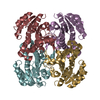
| ||||||||
| Unit cell |
|
-Components
| #1: Protein | Mass: 31530.762 Da / Num. of mol.: 8 Source method: isolated from a genetically manipulated source Details: Expressed with a N-terminal hexahis tag and 3C protease cleavage site but crystallized protein was not cleaved Source: (gene. exp.) Brucella melitensis (bacteria) / Strain: biovar Abortus 2308 / Gene: BMEI1512 / Production host: References: UniProt: Q8YFK8, enoyl-[acyl-carrier-protein] reductase (NADH) #2: Water | ChemComp-HOH / |  Mass: 18.015 Da / Num. of mol.: 563 / Source method: isolated from a natural source / Formula: H2O Mass: 18.015 Da / Num. of mol.: 563 / Source method: isolated from a natural source / Formula: H2O |
|---|
-Experimental details
-Experiment
| Experiment | Method: X-RAY DIFFRACTION / Number of used crystals: 1 |
|---|
- Sample preparation
| Crystal | Density Matthews: 2.14 Å3/Da / Density % sol: 42.49 % |
|---|---|
| Crystal grow | Temperature: 289 K / Method: vapor diffusion, sitting drop / pH: 7 Details: PACT Premier screen condition C11, 20% PEG 6000, 0.2 M CaCl2, 0.1 M HEPES NaOH pH 7.0, 47.3 mg/mL protein, crystal ID 200296c11, VAPOR DIFFUSION, SITTING DROP, temperature 289K |
-Data collection
| Diffraction | Mean temperature: 100 K |
|---|---|
| Diffraction source | Source: ROTATING ANODE / Type: RIGAKU FR-E+ SUPERBRIGHT / Wavelength: 1.5418 Å |
| Detector | Type: RIGAKU SATURN 944 / Detector: CCD / Date: Oct 2, 2008 |
| Radiation | Protocol: SINGLE WAVELENGTH / Monochromatic (M) / Laue (L): M / Scattering type: x-ray |
| Radiation wavelength | Wavelength: 1.5418 Å / Relative weight: 1 |
| Reflection | Resolution: 2.35→20 Å / Num. obs: 84019 / % possible obs: 95.1 % / Observed criterion σ(I): -3 / Biso Wilson estimate: 37.157 Å2 / Rmerge(I) obs: 0.074 / Net I/σ(I): 14.3 |
| Reflection shell | Resolution: 2.35→2.41 Å / Rmerge(I) obs: 0.364 / Mean I/σ(I) obs: 3.6 / Num. measured obs: 16977 / Num. unique all: 5125 / Num. unique obs: 5125 / % possible all: 79.1 |
-Phasing
| Phasing | Method: molecular replacement |
|---|
- Processing
| Software |
| |||||||||||||||||||||||||||||||||||||||||||||||||||||||||||||||||||||||||||||||||||||
|---|---|---|---|---|---|---|---|---|---|---|---|---|---|---|---|---|---|---|---|---|---|---|---|---|---|---|---|---|---|---|---|---|---|---|---|---|---|---|---|---|---|---|---|---|---|---|---|---|---|---|---|---|---|---|---|---|---|---|---|---|---|---|---|---|---|---|---|---|---|---|---|---|---|---|---|---|---|---|---|---|---|---|---|---|---|---|
| Refinement | Method to determine structure: MOLECULAR REPLACEMENT / Resolution: 2.35→19.81 Å / Cor.coef. Fo:Fc: 0.941 / Cor.coef. Fo:Fc free: 0.909 / WRfactor Rfree: 0.212 / WRfactor Rwork: 0.181 / Occupancy max: 1 / Occupancy min: 1 / FOM work R set: 0.833 / SU B: 8.008 / SU ML: 0.192 / SU R Cruickshank DPI: 0.48 / SU Rfree: 0.261 / Cross valid method: THROUGHOUT / σ(F): 0 / ESU R: 0.479 / ESU R Free: 0.289 / Stereochemistry target values: MAXIMUM LIKELIHOOD Details: HYDROGENS HAVE BEEN ADDED IN THE RIDING POSITIONS. U VALUES: REFINED INDIVIDUALLY
| |||||||||||||||||||||||||||||||||||||||||||||||||||||||||||||||||||||||||||||||||||||
| Solvent computation | Ion probe radii: 0.8 Å / Shrinkage radii: 0.8 Å / VDW probe radii: 1.4 Å / Solvent model: MASK | |||||||||||||||||||||||||||||||||||||||||||||||||||||||||||||||||||||||||||||||||||||
| Displacement parameters | Biso max: 62.06 Å2 / Biso mean: 27.873 Å2 / Biso min: 5.39 Å2
| |||||||||||||||||||||||||||||||||||||||||||||||||||||||||||||||||||||||||||||||||||||
| Refinement step | Cycle: LAST / Resolution: 2.35→19.81 Å
| |||||||||||||||||||||||||||||||||||||||||||||||||||||||||||||||||||||||||||||||||||||
| Refine LS restraints |
| |||||||||||||||||||||||||||||||||||||||||||||||||||||||||||||||||||||||||||||||||||||
| LS refinement shell | Resolution: 2.35→2.41 Å / Total num. of bins used: 20
|
