 Movie
Movie Controller


+ Open data

- Basic information
| Entry | Database: PDB / ID: 1iy8 | ||||||
|---|---|---|---|---|---|---|---|
| Title | Crystal Structure of Levodione Reductase | ||||||
 Components Components | LEVODIONE REDUCTASE | ||||||
 Keywords Keywords | OXIDOREDUCTASE | ||||||
| Function / homology |  Function and homology information Function and homology informationOxidoreductases; Acting on the CH-OH group of donors; With NAD+ or NADP+ as acceptor / oxidoreductase activity Similarity search - Function | ||||||
| Biological species |  Leifsonia aquatica (bacteria) Leifsonia aquatica (bacteria) | ||||||
| Method |  X-RAY DIFFRACTION / SYNCHROTRON / MOLECULAR REPLACEMENT / Resolution: 1.6 Å X-RAY DIFFRACTION / SYNCHROTRON / MOLECULAR REPLACEMENT / Resolution: 1.6 Å | ||||||
 Authors Authors | Sogabe, S. / Fukami, T. / Shiratori, Y. / Yoshizumi, A. / Wada, M. | ||||||
 Citation Citation | Journal: J.BIOL.CHEM. / Year: 2003 Title: The Crystal Structure and Stereospecificity of Levodione Reductase from Corynebacterium aquaticum M-13 Authors: Sogabe, S. / Yoshizumi, A. / Fukami, T. / Shiratori, Y. / Shimizu, S. / Takagi, H. / Nakamori, S. / Wada, M. | ||||||
| History |
|
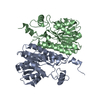
- Structure visualization
| Structure viewer | Molecule: MolmilJmol/JSmol |
|---|
- Downloads & links
-Download
| PDBx/mmCIF format | 1iy8.cif.gz | 402.1 KB | Display | PDBx/mmCIF format |
|---|---|---|---|---|
| PDB format | pdb1iy8.ent.gz | 328.5 KB | Display | PDB format |
| PDBx/mmJSON format | 1iy8.json.gz | Tree view | PDBx/mmJSON format | |
| Others |  Other downloads Other downloads |
-Validation report
| Arichive directory | https://data.pdbj.org/pub/pdb/validation_reports/iy/1iy8ftp://data.pdbj.org/pub/pdb/validation_reports/iy/1iy8 | HTTPS FTP |
|---|
-Related structure data
| Related structure data |  2hsdS S: Starting model for refinement |
|---|---|
| Similar structure data |


-Links
 PDBj
PDBj

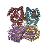
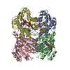
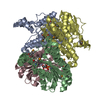
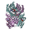
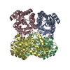
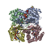
- Assembly
| Deposited unit | 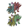
| ||||||||
|---|---|---|---|---|---|---|---|---|---|
| 1 | 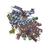
| ||||||||
| 2 | 
| ||||||||
| Unit cell |
|
-Components
| #1: Protein | Mass: 27945.305 Da / Num. of mol.: 8 Source method: isolated from a genetically manipulated source Source: (gene. exp.) Leifsonia aquatica (bacteria) / Strain: M-13 / Plasmid: pkk223-3 / Production host: #2: Chemical | ChemComp-NAD /   Mass: 663.425 Da / Num. of mol.: 8 / Source method: obtained synthetically / Formula: C21H27N7O14P2 / Comment: NAD*YM Mass: 663.425 Da / Num. of mol.: 8 / Source method: obtained synthetically / Formula: C21H27N7O14P2 / Comment: NAD*YM#3: Chemical | ChemComp-MRD / (   Mass: 118.174 Da / Num. of mol.: 8 / Source method: obtained synthetically / Formula: C6H14O2 / Comment: precipitant*YM Mass: 118.174 Da / Num. of mol.: 8 / Source method: obtained synthetically / Formula: C6H14O2 / Comment: precipitant*YM#4: Water | ChemComp-HOH / |  Mass: 18.015 Da / Num. of mol.: 954 / Source method: isolated from a natural source / Formula: H2O Mass: 18.015 Da / Num. of mol.: 954 / Source method: isolated from a natural source / Formula: H2O |
|---|
-Experimental details
-Experiment
| Experiment | Method: X-RAY DIFFRACTION / Number of used crystals: 1 |
|---|
- Sample preparation
| Crystal | Density Matthews: 2.48 Å3/Da / Density % sol: 50.35 % | |||||||||||||||||||||||||||||||||||||||||||||||||
|---|---|---|---|---|---|---|---|---|---|---|---|---|---|---|---|---|---|---|---|---|---|---|---|---|---|---|---|---|---|---|---|---|---|---|---|---|---|---|---|---|---|---|---|---|---|---|---|---|---|---|
| Crystal grow | Temperature: 288 K / Method: vapor diffusion, hanging drop / pH: 6 Details: PEG MME 2000, magnesium chloride, MPD, glycerol, pH 6.0, VAPOR DIFFUSION, HANGING DROP, temperature 288K | |||||||||||||||||||||||||||||||||||||||||||||||||
| Crystal grow | *PLUS Temperature: 15 ℃ / Method: vapor diffusion, hanging drop | |||||||||||||||||||||||||||||||||||||||||||||||||
| Components of the solutions | *PLUS
|
-Data collection
| Diffraction |
| |||||||||||||||
|---|---|---|---|---|---|---|---|---|---|---|---|---|---|---|---|---|
| Diffraction source |
| |||||||||||||||
| Detector |
| |||||||||||||||
| Radiation | Protocol: SINGLE WAVELENGTH / Monochromatic (M) / Laue (L): M / Scattering type: x-ray | |||||||||||||||
| Radiation wavelength |
| |||||||||||||||
| Reflection | Resolution: 1.6→40 Å / Num. obs: 261092 / % possible obs: 92 % / Observed criterion σ(F): 0 / Observed criterion σ(I): 0 | |||||||||||||||
| Reflection shell | Resolution: 1.6→1.7 Å / % possible all: 83.2 | |||||||||||||||
| Reflection | *PLUS Lowest resolution: 50 Å / % possible obs: 92.1 % / Redundancy: 4 % / Num. measured all: 1031771 / Rmerge(I) obs: 0.081 | |||||||||||||||
| Reflection shell | *PLUS % possible obs: 82.7 % / Rmerge(I) obs: 0.263 |

- Processing
| Software |
| ||||||||||||||||||
|---|---|---|---|---|---|---|---|---|---|---|---|---|---|---|---|---|---|---|---|
| Refinement | Method to determine structure: MOLECULAR REPLACEMENT Starting model: PDB ENTRY 2HSD Resolution: 1.6→40 Å / σ(F): 0 / Stereochemistry target values: Engh & Huber
| ||||||||||||||||||
| Refine analyze | Luzzati coordinate error obs: 0.18 Å / Luzzati d res low obs: 5 Å / Luzzati sigma a obs: 0.1 Å | ||||||||||||||||||
| Refinement step | Cycle: LAST / Resolution: 1.6→40 Å
| ||||||||||||||||||
| Refine LS restraints |
| ||||||||||||||||||
| Refinement | *PLUS Num. reflection obs: 16602 / % reflection Rfree: 5 % / Rfactor Rfree: 0.218 | ||||||||||||||||||
| Solvent computation | *PLUS | ||||||||||||||||||
| Displacement parameters | *PLUS | ||||||||||||||||||
| Refine LS restraints | *PLUS Type: c_angle_deg / Dev ideal: 1.534 | ||||||||||||||||||
| LS refinement shell | *PLUS Highest resolution: 1.6 Å / Lowest resolution: 1.7 Å / Rfactor Rfree: 0.281 / Rfactor Rwork: 0.254 |
