 Movie
Movie Controller

[English] 日本語
 Yorodumi
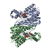
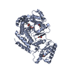
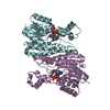
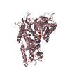
- PDB-3qwi: Crystal structure of a 17beta-hydroxysteroid dehydrogenase (holo ... -
+ Open data

- Basic information
| Entry | Database: PDB / ID: 3qwi | ||||||
|---|---|---|---|---|---|---|---|
| Title | Crystal structure of a 17beta-hydroxysteroid dehydrogenase (holo form) from fungus Cochliobolus lunatus in complex with NADPH and coumestrol | ||||||
 Components Components | 17beta-hydroxysteroid dehydrogenase | ||||||
 Keywords Keywords | OXIDOREDUCTASE/OXIDOREDUCTASE INHIBITOR / 17BETA-HYDROXYSTEROID DEHYDROGENASE / SHORT CHAIN DEHYDROGENASE/REDUCTASE / PHYTOESTROGENS / FLAVONOID / Rossmann Fold / Cytosol / OXIDOREDUCTASE-OXIDOREDUCTASE INHIBITOR complex | ||||||
| Function / homology |  Function and homology information Function and homology informationoxidoreductase activity, acting on CH-OH group of donors / nucleotide binding Similarity search - Function | ||||||
| Biological species |  Cochliobolus lunatus (fungus) Cochliobolus lunatus (fungus) | ||||||
| Method |  X-RAY DIFFRACTION / SYNCHROTRON / MOLECULAR REPLACEMENT / Resolution: 2.5 Å X-RAY DIFFRACTION / SYNCHROTRON / MOLECULAR REPLACEMENT / Resolution: 2.5 Å | ||||||
 Authors Authors | Cassetta, A. / Lamba, D. / Krastanova, I. / Stojan, J. / Lanisnik Rizner, T. / Kristan, K. / Brunskole, M. | ||||||
 Citation Citation | Journal: Biochem.J. / Year: 2012 Title: Structural studies on the flavonoid inhibition of a fungal 17Beta-Hydroxysteroid dehydrogenase Authors: Cassetta, A. / Krastanova, I. / Kristan, K. / Brunskole Svegelj, M. / Lamba, D. / Lanisnik Rizner, T. / Stojan, J. | ||||||
| History |
|
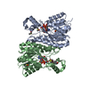
- Structure visualization
| Structure viewer | Molecule: MolmilJmol/JSmol |
|---|
- Downloads & links
-Download
| PDBx/mmCIF format | 3qwi.cif.gz | 406.5 KB | Display | PDBx/mmCIF format |
|---|---|---|---|---|
| PDB format | pdb3qwi.ent.gz | 336 KB | Display | PDB format |
| PDBx/mmJSON format | 3qwi.json.gz | Tree view | PDBx/mmJSON format | |
| Others |  Other downloads Other downloads |
-Validation report
| Arichive directory | https://data.pdbj.org/pub/pdb/validation_reports/qw/3qwiftp://data.pdbj.org/pub/pdb/validation_reports/qw/3qwi | HTTPS FTP |
|---|
-Related structure data
| Related structure data | 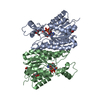 3qwfSC S: Starting model for refinement C: citing same article ( |
|---|---|
| Similar structure data |


-Links
 PDBj
PDBj


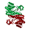
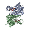
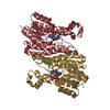
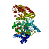
- Assembly
| Deposited unit | 
| ||||||||
|---|---|---|---|---|---|---|---|---|---|
| 1 | 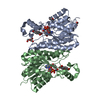
| ||||||||
| 2 | 
| ||||||||
| Unit cell |
|
-Components
| #1: Protein | Mass: 28936.545 Da / Num. of mol.: 4 Source method: isolated from a genetically manipulated source Source: (gene. exp.) Cochliobolus lunatus (fungus) / Strain: M118 / Gene: 17HSDcl / Plasmid: PGEX / Production host:  References: UniProt: O93874, 17beta-estradiol 17-dehydrogenase #2: Chemical | ChemComp-NAP /   Mass: 743.405 Da / Num. of mol.: 4 / Source method: obtained synthetically / Formula: C21H28N7O17P3 Mass: 743.405 Da / Num. of mol.: 4 / Source method: obtained synthetically / Formula: C21H28N7O17P3#3: Chemical |   Mass: 268.221 Da / Num. of mol.: 3 / Source method: obtained synthetically / Formula: C15H8O5 Mass: 268.221 Da / Num. of mol.: 3 / Source method: obtained synthetically / Formula: C15H8O5#4: Chemical | ChemComp-EDO /   Mass: 62.068 Da / Num. of mol.: 9 / Source method: obtained synthetically / Formula: C2H6O2 Mass: 62.068 Da / Num. of mol.: 9 / Source method: obtained synthetically / Formula: C2H6O2#5: Water | ChemComp-HOH / |  Mass: 18.015 Da / Num. of mol.: 329 / Source method: isolated from a natural source / Formula: H2O Mass: 18.015 Da / Num. of mol.: 329 / Source method: isolated from a natural source / Formula: H2O |
|---|
-Experimental details
-Experiment
| Experiment | Method: X-RAY DIFFRACTION / Number of used crystals: 1 |
|---|
- Sample preparation
| Crystal | Density Matthews: 2.19 Å3/Da / Density % sol: 43.95 % |
|---|---|
| Crystal grow | Temperature: 293 K / Method: vapor diffusion, hanging drop / pH: 7.9 Details: 30% (W/V) PEG 2000 MME, 0.1M KCNS, 15% (V/V) Crystals soaked in: 30% (W/V) PEG 2000 MME, 0.1M KCNS, 15% (V/V) ETHYLENE GLYCOLE, 5% (V/V) DMSO, 2MM COUMESTROL, PH 7.9, VAPOR DIFFUSION, ...Details: 30% (W/V) PEG 2000 MME, 0.1M KCNS, 15% (V/V) Crystals soaked in: 30% (W/V) PEG 2000 MME, 0.1M KCNS, 15% (V/V) ETHYLENE GLYCOLE, 5% (V/V) DMSO, 2MM COUMESTROL, PH 7.9, VAPOR DIFFUSION, HANGING DROP, TEMPERATURE 293K |
-Data collection
| Diffraction | Mean temperature: 100 K |
|---|---|
| Diffraction source | Source: SYNCHROTRON / Site: ELETTRA  / Beamline: 5.2R / Wavelength: 1 / Wavelength: 1 Å / Beamline: 5.2R / Wavelength: 1 / Wavelength: 1 Å |
| Detector | Type: MAR CCD 165 mm / Detector: CCD / Date: Aug 5, 2008 / Details: PT COATED TOROIDAL MIRROR |
| Radiation | Monochromator: SI (111) DOUBLE CRYSTAL / Protocol: SINGLE WAVELENGTH / Monochromatic (M) / Laue (L): M / Scattering type: x-ray |
| Radiation wavelength | Wavelength: 1 Å / Relative weight: 1 |
| Reflection | Resolution: 2.5→34.29 Å / Num. all: 34093 / Num. obs: 34093 / % possible obs: 98 % / Observed criterion σ(F): 0 / Observed criterion σ(I): -3 / Redundancy: 3.4 % / Biso Wilson estimate: 40.3 Å2 / Rmerge(I) obs: 0.078 / Rsym value: 0.078 / Net I/σ(I): 11.3 |
| Reflection shell | Resolution: 2.5→2.63 Å / Redundancy: 2.8 % / Rmerge(I) obs: 0.347 / Mean I/σ(I) obs: 3 / Num. unique all: 12947 / Rsym value: 0.347 / % possible all: 91.6 |
- Processing
| Software |
| |||||||||||||||||||||||||||||||||||||||||||||||||||||||||||||||||||||||||||||||||||||||||||||||||||||||||||||||||||||||||||||
|---|---|---|---|---|---|---|---|---|---|---|---|---|---|---|---|---|---|---|---|---|---|---|---|---|---|---|---|---|---|---|---|---|---|---|---|---|---|---|---|---|---|---|---|---|---|---|---|---|---|---|---|---|---|---|---|---|---|---|---|---|---|---|---|---|---|---|---|---|---|---|---|---|---|---|---|---|---|---|---|---|---|---|---|---|---|---|---|---|---|---|---|---|---|---|---|---|---|---|---|---|---|---|---|---|---|---|---|---|---|---|---|---|---|---|---|---|---|---|---|---|---|---|---|---|---|---|
| Refinement | Method to determine structure: MOLECULAR REPLACEMENT Starting model: pdb entry 3QWF Resolution: 2.5→33.86 Å / Cor.coef. Fo:Fc: 0.953 / Cor.coef. Fo:Fc free: 0.895 / SU B: 20.507 / SU ML: 0.207 / Cross valid method: THROUGHOUT / σ(F): 0 / σ(I): 0 / ESU R Free: 0.304 / Stereochemistry target values: MAXIMUM LIKELIHOOD / Details: HYDROGENS HAVE BEEN ADDED IN THE RIDING POSITIONS
| |||||||||||||||||||||||||||||||||||||||||||||||||||||||||||||||||||||||||||||||||||||||||||||||||||||||||||||||||||||||||||||
| Solvent computation | Ion probe radii: 0.8 Å / Shrinkage radii: 0.8 Å / VDW probe radii: 1.4 Å / Solvent model: BABINET MODEL WITH MASK | |||||||||||||||||||||||||||||||||||||||||||||||||||||||||||||||||||||||||||||||||||||||||||||||||||||||||||||||||||||||||||||
| Displacement parameters | Biso mean: 30.013 Å2
| |||||||||||||||||||||||||||||||||||||||||||||||||||||||||||||||||||||||||||||||||||||||||||||||||||||||||||||||||||||||||||||
| Refine analyze | Luzzati coordinate error obs: 0.278 Å | |||||||||||||||||||||||||||||||||||||||||||||||||||||||||||||||||||||||||||||||||||||||||||||||||||||||||||||||||||||||||||||
| Refinement step | Cycle: LAST / Resolution: 2.5→33.86 Å
| |||||||||||||||||||||||||||||||||||||||||||||||||||||||||||||||||||||||||||||||||||||||||||||||||||||||||||||||||||||||||||||
| Refine LS restraints |
| |||||||||||||||||||||||||||||||||||||||||||||||||||||||||||||||||||||||||||||||||||||||||||||||||||||||||||||||||||||||||||||
| LS refinement shell | Resolution: 2.5→2.56 Å / Total num. of bins used: 20
| |||||||||||||||||||||||||||||||||||||||||||||||||||||||||||||||||||||||||||||||||||||||||||||||||||||||||||||||||||||||||||||
| Refinement TLS params. | Method: refined / Refine-ID: X-RAY DIFFRACTION
| |||||||||||||||||||||||||||||||||||||||||||||||||||||||||||||||||||||||||||||||||||||||||||||||||||||||||||||||||||||||||||||
| Refinement TLS group |
|
