coagulation factor Xa / Defective factor IX causes thrombophilia / Defective cofactor function of FVIIIa variant / Defective F9 variant does not activate FX / : / positive regulation of TOR signaling / Transport of gamma-carboxylated protein precursors from the endoplasmic reticulum to the Golgi apparatus / : / Gamma-carboxylation of protein precursors / Removal of aminoterminal propeptides from gamma-carboxylated proteins ...coagulation factor Xa / Defective factor IX causes thrombophilia / Defective cofactor function of FVIIIa variant / Defective F9 variant does not activate FX / : / positive regulation of TOR signaling / Transport of gamma-carboxylated protein precursors from the endoplasmic reticulum to the Golgi apparatus / : / Gamma-carboxylation of protein precursors / Removal of aminoterminal propeptides from gamma-carboxylated proteins / : / phospholipid binding / Golgi lumen / blood coagulation / positive regulation of cell migration / endoplasmic reticulum lumen / serine-type endopeptidase activity / external side of plasma membrane / calcium ion binding / proteolysis / : / extracellular region / plasma membrane Similarity search - Function
SHEET DETERMINATION METHOD: DSSP THE SHEETS PRESENTED AS "AB" IN EACH CHAIN ON SHEET RECORDS BELOW ... SHEET DETERMINATION METHOD: DSSP THE SHEETS PRESENTED AS "AB" IN EACH CHAIN ON SHEET RECORDS BELOW IS ACTUALLY AN 6-STRANDED BARREL THIS IS REPRESENTED BY A 7-STRANDED SHEET IN WHICH THE FIRST AND LAST STRANDS ARE IDENTICAL.
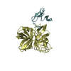
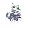
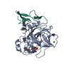
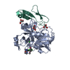
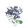
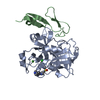
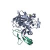
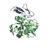
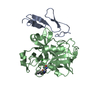
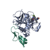
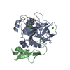
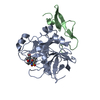
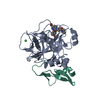
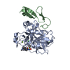
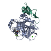
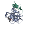
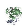
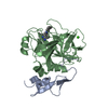
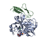
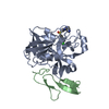
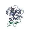
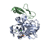
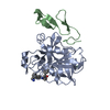
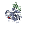
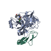
ACTIVATEDFACTORXAHEAVYCHAIN / COAGULATION FACTOR X / STUART FACTOR / STUART-PROWER FACTOR
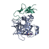
Mass: 28550.596 Da / Num. of mol.: 1 / Source method: isolated from a natural source Details: PURCHASED FROM ENZYME RESEARCH LABS. ISOLATED FROM HUMAN BLOOD Source: (natural) HOMO SAPIENS (human) / References: UniProt: P00742, coagulation factor Xa
#2: Protein
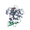
FACTORXLIGHTCHAIN / COAGULATION FACTOR X / STUART FACTOR / STUART-PROWER FACTOR
Mass: 15210.793 Da / Num. of mol.: 1 / Fragment: ACTIVATED DESGLA, RESIDUES 46-179 / Source method: isolated from a natural source Details: PURCHASED FROM ENZYME RESEARCH LABS. ISOLATED FROM HUMAN BLOOD Source: (natural) HOMO SAPIENS (human) / References: UniProt: P00742, coagulation factor Xa
Mass: 18.015 Da / Num. of mol.: 272 / Source method: isolated from a natural source / Formula: H2O
-
Details
Has protein modification
Y
Sequence details
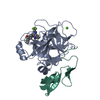
SOME RESIDUES IN CHAIN ARE NOT SEEN IN THE ELECTRON DENSITY. SEQUENCE DATABASE RESIDUES 1-45 (THE ...SOME RESIDUES IN CHAIN ARE NOT SEEN IN THE ELECTRON DENSITY. SEQUENCE DATABASE RESIDUES 1-45 (THE GLA DOMAIN) WERE BIOCHEMICALLY REMOVED IN CHAIN B
-
Experimental details
-
Experiment
Experiment
Method: X-RAY DIFFRACTION / Number of used crystals: 1
-
Sample preparation
Crystal
Density Matthews: 1.81 Å3/Da / Density % sol: 49.3 % / Description: NONE
Crystal grow
Method: vapor diffusion / pH: 5.75 Details: CRYSTALLISATION WAS CARRIED OUT USING VAPOUR DIFFUSION IN 2UL DROPS CONTAINING A 1:1 MIXTURE OF PROTEIN AND WELL SOLUTION. WELL SOLUTION CONTAINED 16-20% PEG 6K, 50 MM MES-NAOH (PH 5.5-6), 5 ...Details: CRYSTALLISATION WAS CARRIED OUT USING VAPOUR DIFFUSION IN 2UL DROPS CONTAINING A 1:1 MIXTURE OF PROTEIN AND WELL SOLUTION. WELL SOLUTION CONTAINED 16-20% PEG 6K, 50 MM MES-NAOH (PH 5.5-6), 5 MM CACL2 AND 50 MM NACL.
Protocol: SINGLE WAVELENGTH / Monochromatic (M) / Laue (L): M / Scattering type: x-ray
Radiation wavelength
Wavelength: 0.961 Å / Relative weight: 1
Reflection
Resolution: 1.69→44.54 Å / Num. obs: 35816 / % possible obs: 97.6 % / Observed criterion σ(I): 0 / Redundancy: 5.5 % / Biso Wilson estimate: 20.615 Å2 / Rmerge(I) obs: 0.08 / Net I/σ(I): 14.4
Reflection shell
Resolution: 1.69→1.78 Å / Redundancy: 5.4 % / Rmerge(I) obs: 0.48 / Mean I/σ(I) obs: 3.4 / % possible all: 100
-
Processing
Software
Name
Version
Classification
REFMAC
5.5.0109
refinement
MOSFLM
datareduction
SCALA
datascaling
Refinement
Method to determine structure: OTHER Starting model: NONE Resolution: 1.7→20 Å / Cor.coef. Fo:Fc: 0.96 / Cor.coef. Fo:Fc free: 0.935 / SU B: 2.119 / SU ML: 0.07 / Cross valid method: THROUGHOUT / ESU R: 0.106 / ESU R Free: 0.107 / Stereochemistry target values: MAXIMUM LIKELIHOOD / Details: HYDROGENS HAVE BEEN ADDED IN THE RIDING POSITIONS.
Rfactor
Num. reflection
% reflection
Selection details
Rfree
0.22473
1753
5 %
RANDOM
Rwork
0.1861
-
-
-
obs
0.18801
33357
100 %
-
Solvent computation
Ion probe radii: 0.8 Å / Shrinkage radii: 0.8 Å / VDW probe radii: 1.4 Å / Solvent model: BABINET MODEL WITH MASK
 Movie
Movie Controller

 Yorodumi
 Open data
 Basic information
 Components
 Keywords
 Function and homology information
 HOMO SAPIENS (human)
 X-RAY DIFFRACTION /
  Authors
 Citation
 Structure visualization
 Downloads & links
 Other downloads


 PDBj
PDBj


 Assembly
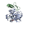
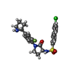


 Mass: 487.974 Da / Num. of mol.: 1 / Source method: obtained synthetically / Formula: C24H23ClFN3O3S
 Mass: 40.078 Da / Num. of mol.: 1 / Source method: obtained synthetically / Formula: Ca
 Mass: 24.305 Da / Num. of mol.: 1 / Source method: obtained synthetically / Formula: Mg
 Sample preparation
 / Beamline: ID29 / Wavelength: 0.961
  Processing