 Movie
Movie Controller

[English] 日本語
 Yorodumi
- PDB-1xkb: FACTOR XA COMPLEXED WITH A SYNTHETIC INHIBITOR FX-2212A,(2S)-(3'-... -
+ Open data

- Basic information
| Entry | Database: PDB / ID: 1xkb | ||||||
|---|---|---|---|---|---|---|---|
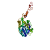
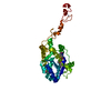
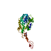
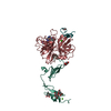
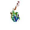
| Title | FACTOR XA COMPLEXED WITH A SYNTHETIC INHIBITOR FX-2212A,(2S)-(3'-AMIDINO-3-BIPHENYLYL)-5-(4-PYRIDYLAMINO)PENTANOIC ACID | ||||||
 Components Components | (BLOOD COAGULATION FACTOR XA) x 2 | ||||||
 Keywords Keywords | BLOOD COAGULATION FACTOR / SERINE PROTEINASE / EPIDERMAL GROWTH FACTOR LIKE DOMAIN | ||||||
| Function / homology |  Function and homology information Function and homology informationcoagulation factor Xa / Defective factor IX causes thrombophilia / Defective cofactor function of FVIIIa variant / Defective F9 variant does not activate FX / : / positive regulation of TOR signaling / Transport of gamma-carboxylated protein precursors from the endoplasmic reticulum to the Golgi apparatus / : / Gamma-carboxylation of protein precursors / Removal of aminoterminal propeptides from gamma-carboxylated proteins ...coagulation factor Xa / Defective factor IX causes thrombophilia / Defective cofactor function of FVIIIa variant / Defective F9 variant does not activate FX / : / positive regulation of TOR signaling / Transport of gamma-carboxylated protein precursors from the endoplasmic reticulum to the Golgi apparatus / : / Gamma-carboxylation of protein precursors / Removal of aminoterminal propeptides from gamma-carboxylated proteins / : / phospholipid binding / Golgi lumen / blood coagulation / positive regulation of cell migration / endoplasmic reticulum lumen / serine-type endopeptidase activity / external side of plasma membrane / calcium ion binding / proteolysis / : / extracellular region / plasma membrane Similarity search - Function | ||||||
| Biological species |  Homo sapiens (human) Homo sapiens (human) | ||||||
| Method |  X-RAY DIFFRACTION / MOLECULAR REPLACEMENT / Resolution: 2.4 Å X-RAY DIFFRACTION / MOLECULAR REPLACEMENT / Resolution: 2.4 Å | ||||||
 Authors Authors | Kamata, K. / Kim, S.H. | ||||||
 Citation Citation | Journal: Proc.Natl.Acad.Sci.USA / Year: 1998 Title: Structural basis for chemical inhibition of human blood coagulation factor Xa. Authors: Kamata, K. / Kawamoto, H. / Honma, T. / Iwama, T. / Kim, S.H. #1: Journal: J.Biol.Chem. / Year: 1996Title: X-Ray Structure of Active Site-Inhibited Clotting Factor Xa. Implications for Drug Design and Substrate Recognition Authors: Brandstetter, H. / Kuhne, A. / Bode, W. / Huber, R. / Von Der Saal, W. / Wirthensohn, K. / Engh, R.A. #2: Journal: J.Mol.Biol. / Year: 1993Title: Structure of Human Des(1-45) Factor Xa at 2.2 A Resolution Authors: Padmanabhan, K. / Padmanabhan, K.P. / Tulinsky, A. / Park, C.H. / Bode, W. / Huber, R. / Blankenship, D.T. / Cardin, A.D. / Kisiel, W. | ||||||
| History |
|
- Structure visualization
| Structure viewer | Molecule: MolmilJmol/JSmol |
|---|
- Downloads & links
-Download
| PDBx/mmCIF format | 1xkb.cif.gz | 147.7 KB | Display | PDBx/mmCIF format |
|---|---|---|---|---|
| PDB format | pdb1xkb.ent.gz | 114.5 KB | Display | PDB format |
| PDBx/mmJSON format | 1xkb.json.gz | Tree view | PDBx/mmJSON format | |
| Others |  Other downloads Other downloads |
-Validation report
| Arichive directory | https://data.pdbj.org/pub/pdb/validation_reports/xk/1xkbftp://data.pdbj.org/pub/pdb/validation_reports/xk/1xkb | HTTPS FTP |
|---|
-Related structure data
| Related structure data |  1xkaC  1hcgS S: Starting model for refinement C: citing same article ( |
|---|---|
| Similar structure data |


-Links
 PDBj
PDBj


- Assembly
| Deposited unit | 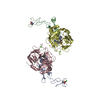
| ||||||||
|---|---|---|---|---|---|---|---|---|---|
| 1 | 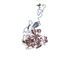
| ||||||||
| 2 | 
| ||||||||
| 3 | 
| ||||||||
| 4 | 
| ||||||||
| Unit cell |
|
-Components
| #1: Protein | Mass: 10386.538 Da / Num. of mol.: 2 / Fragment: PROTEOLYTIC CLEAVAGE PRODUCT, GLA DOMAIN / Source method: isolated from a natural source / Source: (natural) Homo sapiens (human) / Tissue: BLOOD / References: UniProt: P00742, coagulation factor Xa#2: Protein | Mass: 26604.297 Da / Num. of mol.: 2 / Fragment: PROTEOLYTIC CLEAVAGE PRODUCT, GLA DOMAIN / Source method: isolated from a natural source / Source: (natural) Homo sapiens (human) / Tissue: BLOOD / References: UniProt: P00742, coagulation factor Xa#3: Chemical |   Mass: 40.078 Da / Num. of mol.: 3 / Source method: obtained synthetically / Formula: Ca Mass: 40.078 Da / Num. of mol.: 3 / Source method: obtained synthetically / Formula: Ca#4: Chemical |   Mass: 388.462 Da / Num. of mol.: 2 / Source method: obtained synthetically / Formula: C23H24N4O2 Mass: 388.462 Da / Num. of mol.: 2 / Source method: obtained synthetically / Formula: C23H24N4O2#5: Water | ChemComp-HOH / |  Mass: 18.015 Da / Num. of mol.: 299 / Source method: isolated from a natural source / Formula: H2O Mass: 18.015 Da / Num. of mol.: 299 / Source method: isolated from a natural source / Formula: H2OHas protein modification | Y | |
|---|
-Experimental details
-Experiment
| Experiment | Method: X-RAY DIFFRACTION / Number of used crystals: 1 |
|---|
- Sample preparation
| Crystal | Density Matthews: 2.5 Å3/Da / Density % sol: 60 % | ||||||||||||||||||||||||||||||||||||||||||
|---|---|---|---|---|---|---|---|---|---|---|---|---|---|---|---|---|---|---|---|---|---|---|---|---|---|---|---|---|---|---|---|---|---|---|---|---|---|---|---|---|---|---|---|
| Crystal grow | pH: 6 / Details: pH 6.0 | ||||||||||||||||||||||||||||||||||||||||||
| Crystal grow | *PLUS Method: vapor diffusion | ||||||||||||||||||||||||||||||||||||||||||
| Components of the solutions | *PLUS
|
-Data collection
| Diffraction | Mean temperature: 100 K |
|---|---|
| Diffraction source | Source: ROTATING ANODE / Type: RIGAKU RUH2R / Wavelength: 1.5418 |
| Detector | Type: RIGAKU / Detector: IMAGE PLATE / Date: Oct 24, 1996 / Details: YALE MIRRORS |
| Radiation | Monochromator: NI FILTER / Monochromatic (M) / Laue (L): M / Scattering type: x-ray |
| Radiation wavelength | Wavelength: 1.5418 Å / Relative weight: 1 |
| Reflection | Resolution: 2.4→30 Å / Num. obs: 26594 / % possible obs: 91.2 % / Observed criterion σ(I): 1 / Redundancy: 2.7 % / Biso Wilson estimate: 46 Å2 / Rmerge(I) obs: 0.057 / Rsym value: 0.057 / Net I/σ(I): 17.7 |
| Reflection shell | Resolution: 2.4→2.53 Å / Redundancy: 1.1 % / Rmerge(I) obs: 0.252 / Mean I/σ(I) obs: 4.1 / Rsym value: 0.252 / % possible all: 58.4 |
| Reflection | *PLUS Num. measured all: 78773 |
| Reflection shell | *PLUS % possible obs: 58.4 % |
- Processing
| Software |
| ||||||||||||||||||||||||||||||||||||||||||||||||||||||||||||||||||||||||||||||||
|---|---|---|---|---|---|---|---|---|---|---|---|---|---|---|---|---|---|---|---|---|---|---|---|---|---|---|---|---|---|---|---|---|---|---|---|---|---|---|---|---|---|---|---|---|---|---|---|---|---|---|---|---|---|---|---|---|---|---|---|---|---|---|---|---|---|---|---|---|---|---|---|---|---|---|---|---|---|---|---|---|---|
| Refinement | Method to determine structure: MOLECULAR REPLACEMENT Starting model: PDB ENTRY 1HCG Resolution: 2.4→8 Å / Rfactor Rfree error: 0.007 / Data cutoff high absF: 1000000 / Data cutoff low absF: 0.001 / Isotropic thermal model: RESTRAINED / Cross valid method: THROUGHOUT / σ(F): 2
| ||||||||||||||||||||||||||||||||||||||||||||||||||||||||||||||||||||||||||||||||
| Displacement parameters | Biso mean: 24.1 Å2 | ||||||||||||||||||||||||||||||||||||||||||||||||||||||||||||||||||||||||||||||||
| Refine analyze |
| ||||||||||||||||||||||||||||||||||||||||||||||||||||||||||||||||||||||||||||||||
| Refinement step | Cycle: LAST / Resolution: 2.4→8 Å
| ||||||||||||||||||||||||||||||||||||||||||||||||||||||||||||||||||||||||||||||||
| Refine LS restraints |
| ||||||||||||||||||||||||||||||||||||||||||||||||||||||||||||||||||||||||||||||||
| LS refinement shell | Resolution: 2.4→2.51 Å / Rfactor Rfree error: 0.036 / Total num. of bins used: 8
| ||||||||||||||||||||||||||||||||||||||||||||||||||||||||||||||||||||||||||||||||
| Xplor file |
| ||||||||||||||||||||||||||||||||||||||||||||||||||||||||||||||||||||||||||||||||
| Software | *PLUS Name: X-PLOR / Version: 3.8 / Classification: refinement | ||||||||||||||||||||||||||||||||||||||||||||||||||||||||||||||||||||||||||||||||
| Refinement | *PLUS | ||||||||||||||||||||||||||||||||||||||||||||||||||||||||||||||||||||||||||||||||
| Solvent computation | *PLUS | ||||||||||||||||||||||||||||||||||||||||||||||||||||||||||||||||||||||||||||||||
| Displacement parameters | *PLUS | ||||||||||||||||||||||||||||||||||||||||||||||||||||||||||||||||||||||||||||||||
| Refine LS restraints | *PLUS
| ||||||||||||||||||||||||||||||||||||||||||||||||||||||||||||||||||||||||||||||||
| LS refinement shell | *PLUS Rfactor obs: 0.354 |
