 Movie
Movie Controller


+ Open data

- Basic information
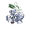
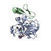
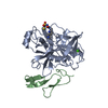
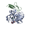
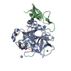
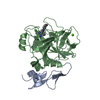
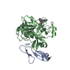
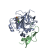
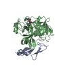
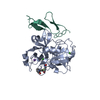
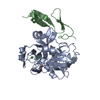
| Entry | Database: PDB / ID: 1iqm | ||||||
|---|---|---|---|---|---|---|---|
| Title | Human coagulation factor Xa in complex with M54471 | ||||||
 Components Components | (coagulation Factor Xa) x 2 | ||||||
 Keywords Keywords | HYDROLASE / SERINE PROTEASE / BLOOD COAGULATION FACTOR / COMPLEX | ||||||
| Function / homology |  Function and homology information Function and homology informationcoagulation factor Xa / Defective factor IX causes thrombophilia / Defective cofactor function of FVIIIa variant / Defective F9 variant does not activate FX / : / positive regulation of TOR signaling / Transport of gamma-carboxylated protein precursors from the endoplasmic reticulum to the Golgi apparatus / : / Gamma-carboxylation of protein precursors / Removal of aminoterminal propeptides from gamma-carboxylated proteins ...coagulation factor Xa / Defective factor IX causes thrombophilia / Defective cofactor function of FVIIIa variant / Defective F9 variant does not activate FX / : / positive regulation of TOR signaling / Transport of gamma-carboxylated protein precursors from the endoplasmic reticulum to the Golgi apparatus / : / Gamma-carboxylation of protein precursors / Removal of aminoterminal propeptides from gamma-carboxylated proteins / : / phospholipid binding / Golgi lumen / blood coagulation / positive regulation of cell migration / endoplasmic reticulum lumen / serine-type endopeptidase activity / external side of plasma membrane / calcium ion binding / proteolysis / : / extracellular region / plasma membrane Similarity search - Function | ||||||
| Biological species |  Homo sapiens (human) Homo sapiens (human) | ||||||
| Method |  X-RAY DIFFRACTION / MOLECULAR REPLACEMENT / Resolution: 2.6 Å X-RAY DIFFRACTION / MOLECULAR REPLACEMENT / Resolution: 2.6 Å | ||||||
 Authors Authors | Shiromizu, I. / Matsusue, T. | ||||||
 Citation Citation | Journal: To be Published Title: Factor Xa Specific Inhibitor that Induces the Novel Binding Model in Complex with Human Fxa Authors: Matsusue, T. / Shiromizu, I. / Okamoto, A. / Nakayama, K. / Nishida, H. / Mukaihira, T. / Miyazaki, Y. / Saitou, F. / Morishita, H. / Ohnishi, S. / Mochizuki, H. #1: Journal: J.Mol.Biol. / Year: 1993Title: Structure of Human Des(1-45) Factor Xa at 2.2 A Resolution Authors: Padmanabhan, K. / Padmanabhan, K.P. / Tulinsky, A. / Park, C.H. / Bode, W. / Huber, R. / Blankenship, D.T. / Cardin, A.D. / Kisiel, W. #2: Journal: J.Biol.Chem. / Year: 1996Title: X-Ray Structure of Active Site-Inhibited Clotting Factor Xa. Implications for Drug Design and Substrate Recognition Authors: Brandstetter, H. / Kuhne, A. / Bode, W. / Huber, R. / Von Der Saal, W. / Wirthensohn, K. / Engh, R.A. #3: Journal: J.Biol.Chem. / Year: 1996Title: Autoproteolysis or Plasmin-Mediated Cleavage of Factor Xaalpha Exposes a Plasminogen Binding Site and Inhibits Coagulation Authors: Pryzdial, E.L. / Kessler, G.E. | ||||||
| History |
|
- Structure visualization
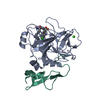
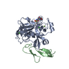
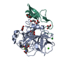
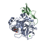
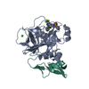
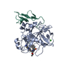
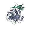
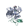
| Structure viewer | Molecule: MolmilJmol/JSmol |
|---|
- Downloads & links
-Download
| PDBx/mmCIF format | 1iqm.cif.gz | 73 KB | Display | PDBx/mmCIF format |
|---|---|---|---|---|
| PDB format | pdb1iqm.ent.gz | 52.6 KB | Display | PDB format |
| PDBx/mmJSON format | 1iqm.json.gz | Tree view | PDBx/mmJSON format | |
| Others |  Other downloads Other downloads |
-Validation report
| Arichive directory | https://data.pdbj.org/pub/pdb/validation_reports/iq/1iqmftp://data.pdbj.org/pub/pdb/validation_reports/iq/1iqm | HTTPS FTP |
|---|
-Related structure data
| Related structure data | 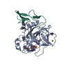 1iqeC  1iqfC  1iqgC  1iqhC  1iqiC  1iqjC  1iqkC  1iqlC  1iqnC C: citing same article ( |
|---|---|
| Similar structure data |


-Links
 PDBj
PDBj


- Assembly
| Deposited unit | 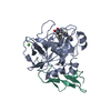
| ||||||||
|---|---|---|---|---|---|---|---|---|---|
| 1 |
| ||||||||
| Unit cell |
|
-Components
| #1: Protein | Mass: 26604.297 Da / Num. of mol.: 1 / Fragment: heavy chain, catalytic domain (residues 235-469) / Source method: isolated from a natural source / Source: (natural) Homo sapiens (human) / References: UniProt: P00742, coagulation factor Xa |
|---|---|
| #2: Protein | Mass: 10533.713 Da / Num. of mol.: 1 Fragment: light chain, epidermal growth factor like domain (residues 84-179) Source method: isolated from a natural source / Source: (natural) Homo sapiens (human) / References: UniProt: P00742, coagulation factor Xa |
| #3: Chemical | ChemComp-CA /   Mass: 40.078 Da / Num. of mol.: 1 / Source method: obtained synthetically / Formula: Ca Mass: 40.078 Da / Num. of mol.: 1 / Source method: obtained synthetically / Formula: Ca |
| #4: Chemical | ChemComp-XMK / 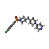  Mass: 461.020 Da / Num. of mol.: 1 / Source method: obtained synthetically / Formula: C23H29ClN4O2S Mass: 461.020 Da / Num. of mol.: 1 / Source method: obtained synthetically / Formula: C23H29ClN4O2S |
| #5: Water | ChemComp-HOH /  Mass: 18.015 Da / Num. of mol.: 40 / Source method: isolated from a natural source / Formula: H2O Mass: 18.015 Da / Num. of mol.: 40 / Source method: isolated from a natural source / Formula: H2O |
| Has protein modification | Y |
-Experimental details
-Experiment
| Experiment | Method: X-RAY DIFFRACTION / Number of used crystals: 1 |
|---|
- Sample preparation
| Crystal | Density Matthews: 2.16 Å3/Da / Density % sol: 43.02 % |
|---|---|
| Crystal grow | Temperature: 293 K / Method: vapor diffusion, sitting drop / pH: 7.2 Details: PEG1500, calcium chloride, M54471, tris-HCl, pH 7.20, VAPOR DIFFUSION, SITTING DROP, temperature 293K |
-Data collection
| Diffraction | Mean temperature: 100 K |
|---|---|
| Diffraction source | Source: ROTATING ANODE / Type: RIGAKU / Wavelength: 1.5418 / Wavelength: 1.5418 Å |
| Detector | Type: RIGAKU RAXIS IV / Detector: IMAGE PLATE / Date: Jun 25, 1999 / Details: MIRRORS |
| Radiation | Monochromator: YALE MIRRORS / Protocol: SINGLE WAVELENGTH / Monochromatic (M) / Laue (L): M / Scattering type: x-ray |
| Radiation wavelength | Wavelength: 1.5418 Å / Relative weight: 1 |
| Reflection | Highest resolution: 2.6 Å / Num. obs: 8545 / % possible obs: 82.2 % / Observed criterion σ(I): 1 / Biso Wilson estimate: 28.3 Å2 / Rmerge(I) obs: 0.054 |
| Reflection shell | Resolution: 2.6→2.7 Å / Rmerge(I) obs: 0.081 / % possible all: 66.4 |
- Processing
| Software |
| ||||||||||||||||||||||||||||||||||||
|---|---|---|---|---|---|---|---|---|---|---|---|---|---|---|---|---|---|---|---|---|---|---|---|---|---|---|---|---|---|---|---|---|---|---|---|---|---|
| Refinement | Method to determine structure: MOLECULAR REPLACEMENT / Resolution: 2.6→8 Å / Rfactor Rfree error: 0.01 / Data cutoff high absF: 16447.98 / Data cutoff low absF: 0 / Isotropic thermal model: RESTRAINED / Cross valid method: THROUGHOUT / σ(F): 0
| ||||||||||||||||||||||||||||||||||||
| Solvent computation | Bsol: 52.6429 Å2 / ksol: 0.502811 e/Å3 | ||||||||||||||||||||||||||||||||||||
| Displacement parameters | Biso mean: 29.2 Å2
| ||||||||||||||||||||||||||||||||||||
| Refine analyze |
| ||||||||||||||||||||||||||||||||||||
| Refinement step | Cycle: LAST / Resolution: 2.6→8 Å
| ||||||||||||||||||||||||||||||||||||
| Refine LS restraints |
| ||||||||||||||||||||||||||||||||||||
| LS refinement shell | Resolution: 2.6→2.76 Å / Rfactor Rfree error: 0.032 / Total num. of bins used: 6
|
