 Movie
Movie Controller

[English] 日本語
 Yorodumi
- PDB-1mq5: Crystal Structure of 3-chloro-N-[4-chloro-2-[[(4-chlorophenyl)ami... -
+ Open data

- Basic information
| Entry | Database: PDB / ID: 1mq5 | ||||||
|---|---|---|---|---|---|---|---|
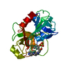
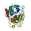
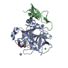
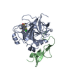
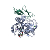
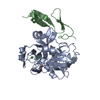
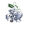
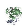
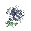
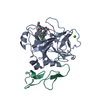
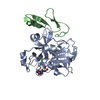
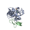
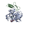
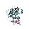
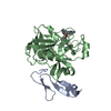
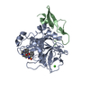
| Title | Crystal Structure of 3-chloro-N-[4-chloro-2-[[(4-chlorophenyl)amino]carbonyl]phenyl]-4-[(4-methyl-1-piperazinyl)methyl]-2-thiophenecarboxamide Complexed with Human Factor Xa | ||||||
 Components Components | (COAGULATION FACTOR X ...) x 2 | ||||||
 Keywords Keywords | BLOOD CLOTTING / PROTEIN INHIBITOR COMPLEX / COAGULATION COFACTOR / PROTEASE | ||||||
| Function / homology |  Function and homology information Function and homology informationcoagulation factor Xa / Defective factor IX causes thrombophilia / Defective cofactor function of FVIIIa variant / Defective F9 variant does not activate FX / : / positive regulation of TOR signaling / Transport of gamma-carboxylated protein precursors from the endoplasmic reticulum to the Golgi apparatus / : / Gamma-carboxylation of protein precursors / Removal of aminoterminal propeptides from gamma-carboxylated proteins ...coagulation factor Xa / Defective factor IX causes thrombophilia / Defective cofactor function of FVIIIa variant / Defective F9 variant does not activate FX / : / positive regulation of TOR signaling / Transport of gamma-carboxylated protein precursors from the endoplasmic reticulum to the Golgi apparatus / : / Gamma-carboxylation of protein precursors / Removal of aminoterminal propeptides from gamma-carboxylated proteins / : / phospholipid binding / Golgi lumen / blood coagulation / positive regulation of cell migration / endoplasmic reticulum lumen / serine-type endopeptidase activity / external side of plasma membrane / calcium ion binding / proteolysis / : / extracellular region / plasma membrane Similarity search - Function | ||||||
| Biological species |  Homo sapiens (human) Homo sapiens (human) | ||||||
| Method |  X-RAY DIFFRACTION / SYNCHROTRON / DIRECT REPLACEMENT / Resolution: 2.1 Å X-RAY DIFFRACTION / SYNCHROTRON / DIRECT REPLACEMENT / Resolution: 2.1 Å | ||||||
 Authors Authors | Adler, M. / Whitlow, M. | ||||||
 Citation Citation | Journal: Biochemistry / Year: 2002 Title: Crystal Structures of Two Potent Nonamidine Inhibitors Bound to Factor Xa Authors: Adler, M. / Kochanny, M.J. / Bin, Y. / Rumennik, G. / Light, D.L. / Biancalana, S. / Whitlow, M. #1: Journal: Biochemistry / Year: 2000Title: Preparation, Characterization and the Crystal Structure of the Inhibitor Zk-807834 (Ci-1031) Complexed with Factor Xa Authors: Adler, M. / Davey, D.D. / Phillips, G.B. / Kim, S.H. / Jancarik, J. / Rumennik, G. / Light, D.L. / Whitlow, M. #2: Journal: J.Med.Chem. / Year: 2000Title: Crystal Structures of Human Factor Xa Complexed with Potent Inhibitors Authors: Maignan, S. / Guilloteau, J.P. / Pouzieux, S. / Choi-Sledeski, Y.M. / Becker, M.R. / Klein, S.I. / Ewing, W.R. / Pauls, H.W. / Spada, A.P. / Mikol, V. #3: Journal: J.Med.Chem. / Year: 1998Title: Discovery of N-[2-[5-[Amino(imino)methyl]-2-hydroxyphenoxy]-3,5-difluoro- 6-[3-(4,5-dihydro-1-methyl-1H-imidazol-2-yl)phenoxy]pyridin-4-yl]-N-methylglycine (ZK-807834): A Potent, Selective, ...Title: Discovery of N-[2-[5-[Amino(imino)methyl]-2-hydroxyphenoxy]-3,5-difluoro- 6-[3-(4,5-dihydro-1-methyl-1H-imidazol-2-yl)phenoxy]pyridin-4-yl]-N-methylglycine (ZK-807834): A Potent, Selective, and Orally Active Inhibitor of the Blood Coagulation Enzyme Factor Xa Authors: Phillips, G.B. / Buckman, B.O. / Davey, D.D. / Eagen, K.A. / Guilford, W.J. / Hinchman, J. / Ho, E. / Koovakkat, S. / Liang, A.M. / Light, D.R. / Mohan, R. / Ng, H.P. / Post, J.M. / Shaw, K. ...Authors: Phillips, G.B. / Buckman, B.O. / Davey, D.D. / Eagen, K.A. / Guilford, W.J. / Hinchman, J. / Ho, E. / Koovakkat, S. / Liang, A.M. / Light, D.R. / Mohan, R. / Ng, H.P. / Post, J.M. / Shaw, K.J. / Smith, D. / Subramanyam, B. / Sullivan, M.E. / Trinh, L. / Vergona, R. / Walters, J. / White, K. / Whitlow, M. / Wu, S. / Xu, W. / Morrissey, M.M. | ||||||
| History |
| ||||||
| Remark 300 | BIOMOLECULE: 1 THIS ENTRY CONTAINS THE CRYSTALLOGRAPHIC ASYMMETRIC UNIT WHICH CONSISTS OF 2 ... BIOMOLECULE: 1 THIS ENTRY CONTAINS THE CRYSTALLOGRAPHIC ASYMMETRIC UNIT WHICH CONSISTS OF 2 CHAIN(S). FACTOR XA FORMS A COMPLEX WITH FACTOR VA IN THE PRESENCE OF CALCIUM AND A PHOSPHOLIPID MEMBRANE TO PRODUCE THE PROTHROMBINASE COMPLEX. THIS ENTRY CONTAINS THE EPIDERMAL GROWTH FACTOR LIKE DOMAIN 2(L) AND THE CATALYTIC DOMAIN (A) OF FACTOR XA IN THE CRYSTALLOGRAPHIC ASYMMETRIC UNIT. THE COORDINATES DO NOT CONTAIN THE GLA DOMAIN OR THE EPIDERMAL GROWTH FACTOR LIKE DOMAIN 1 OF FACTOR XA. ALTHOUGH THE ASYMMETRIC UNIT CONTAINS A FUNCTIONAL PROTEASE, IT DOES HAVE THE SAME SPECIFICITY AS THE PROTHROMBINASE COMPLEX. SEE REMARK 350 FOR INFORMATION ON GENERATING THE BIOLOGICAL MOLECULE(S). |
- Structure visualization
| Structure viewer | Molecule: MolmilJmol/JSmol |
|---|
- Downloads & links
-Download
| PDBx/mmCIF format | 1mq5.cif.gz | 78.1 KB | Display | PDBx/mmCIF format |
|---|---|---|---|---|
| PDB format | pdb1mq5.ent.gz | 56.1 KB | Display | PDB format |
| PDBx/mmJSON format | 1mq5.json.gz | Tree view | PDBx/mmJSON format | |
| Others |  Other downloads Other downloads |
-Validation report
| Arichive directory | https://data.pdbj.org/pub/pdb/validation_reports/mq/1mq5ftp://data.pdbj.org/pub/pdb/validation_reports/mq/1mq5 | HTTPS FTP |
|---|
-Related structure data
| Related structure data | 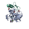 1mq6C  1fjsS S: Starting model for refinement C: citing same article ( |
|---|---|
| Similar structure data |


-Links
 PDBj
PDBj


- Assembly
| Deposited unit | 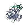
| ||||||||
|---|---|---|---|---|---|---|---|---|---|
| 1 |
| ||||||||
| Unit cell |
| ||||||||
| Details | Factor Xa forms a complex with factor Va in the presence of calcium and a phospholipid membrane to produce the prothrombinase complex. |
-Components
-COAGULATION FACTOR X ... , 2 types, 2 molecules AL
| #1: Protein | Mass: 26346.000 Da / Num. of mol.: 1 / Fragment: CATALYTIC DOMAIN / Source method: isolated from a natural source / Details: EXTRACTED FROM BLOOD / Source: (natural) Homo sapiens (human) / References: UniProt: P00742, coagulation factor Xa |
|---|---|
| #2: Protein | Mass: 5460.121 Da / Num. of mol.: 1 / Fragment: EPIDERMAL GROWTH FACTOR LIKE DOMAIN 2 / Source method: isolated from a natural source / Details: EXTRACTED FROM BLOOD / Source: (natural) Homo sapiens (human) / References: UniProt: P00742, coagulation factor Xa |
-Non-polymers , 4 types, 177 molecules 


| #3: Chemical | ChemComp-CA /  Mass: 40.078 Da / Num. of mol.: 1 / Source method: obtained synthetically / Formula: Ca Mass: 40.078 Da / Num. of mol.: 1 / Source method: obtained synthetically / Formula: Ca | ||
|---|---|---|---|
| #4: Chemical | ChemComp-XLC /  Mass: 537.889 Da / Num. of mol.: 1 / Source method: obtained synthetically / Formula: C24H23Cl3N4O2S Mass: 537.889 Da / Num. of mol.: 1 / Source method: obtained synthetically / Formula: C24H23Cl3N4O2S | ||
| #5: Chemical |  Mass: 92.094 Da / Num. of mol.: 3 / Source method: obtained synthetically / Formula: C3H8O3 Mass: 92.094 Da / Num. of mol.: 3 / Source method: obtained synthetically / Formula: C3H8O3#6: Water | ChemComp-HOH / | Mass: 18.015 Da / Num. of mol.: 172 / Source method: isolated from a natural source / Formula: H2O |
-Details
| Has protein modification | Y |
|---|
-Experimental details
-Experiment
| Experiment | Method: X-RAY DIFFRACTION / Number of used crystals: 1 |
|---|
- Sample preparation
| Crystal | Density Matthews: 2.44 Å3/Da / Density % sol: 51.2 % | ||||||||||||||||||||||||||||||||||||||||||
|---|---|---|---|---|---|---|---|---|---|---|---|---|---|---|---|---|---|---|---|---|---|---|---|---|---|---|---|---|---|---|---|---|---|---|---|---|---|---|---|---|---|---|---|
| Crystal grow | Temperature: 293 K / Method: vapor diffusion, hanging drop / pH: 7.5 Details: A THREE-FOLD EXCESS OF A PROPRIETARY FACTOR XA INHIBITOR WITH PICOMOLAR AFFINITY WAS ADDED TO THE DES-GLA-FACTOR XA. THIS INHIBITOR IS BASED ON THE SAME TEMPLATE AS XLC IN 1MQ5. THE PROTEIN ...Details: A THREE-FOLD EXCESS OF A PROPRIETARY FACTOR XA INHIBITOR WITH PICOMOLAR AFFINITY WAS ADDED TO THE DES-GLA-FACTOR XA. THIS INHIBITOR IS BASED ON THE SAME TEMPLATE AS XLC IN 1MQ5. THE PROTEIN WAS THEN CONCENTRATED TO 12-17 MG/ML. CRYSTALS WERE GROWN USING 2 UL OF COMPLEX WITH 2 UL OF RESERVOIR CONTAINING 15-21% PEG1500 AND 10 MM CACL2. 30-40 UL SITTING DROPS CONTAINING SATURATED INHIBITOR (5 MM) IN 21% PEG1500, 5 MM CACL2, 50 MM NACL, 50 MM TRIS PH 7.5 (CRYSTAL SOAKING SOLUTION) WERE EQUILIBRATED OVER A 1 ML RESERVOIR CONTAINING THE CRYSTAL SOAKING SOLUTION FOR 1 TO 2 DAYS. A SINGLE FACTOR XA CRYSTAL WAS TRANSFERRED USING A MOUNTED CRYOLOOP INTO ONE OF THESE SITTING DROPS AND ALLOWED TO SOAK FOR THREE OR MORE Days. AFTER THE INITIAL SOAK, EACH CRYSTAL WAS THEN TRANSFERRED TO AN UNUSED DROP AND ALLOWED TO SOAK FOR SECOND PERIOD OF THREE OR MORE DAYS. pH 7.50, VAPOR DIFFUSION, HANGING DROP, temperature 293K | ||||||||||||||||||||||||||||||||||||||||||
| Crystal grow | *PLUS Details: Adler, M., (2000) Biochemistry, 39, 12534. | ||||||||||||||||||||||||||||||||||||||||||
| Components of the solutions | *PLUS
|
-Data collection
| Diffraction | Mean temperature: 100 K | |||||||||
|---|---|---|---|---|---|---|---|---|---|---|
| Diffraction source | Source: SYNCHROTRON / Site: SSRL  / Beamline: BL7-1 / Wavelength: 1.08 / Wavelength: 1.08 Å / Beamline: BL7-1 / Wavelength: 1.08 / Wavelength: 1.08 Å | |||||||||
| Detector | Type: MARRESEARCH / Detector: IMAGE PLATE / Date: Dec 6, 2000 | |||||||||
| Radiation | Monochromator: SI(111) / Protocol: SINGLE WAVELENGTH / Monochromatic (M) / Laue (L): M / Scattering type: x-ray | |||||||||
| Radiation wavelength |
| |||||||||
| Reflection | Resolution: 2.1→25 Å / Num. all: 18977 / Num. obs: 18977 / % possible obs: 98.4 % / Observed criterion σ(F): 0 / Observed criterion σ(I): 0 / Redundancy: 3.6 % / Biso Wilson estimate: 30.79 Å2 / Rsym value: 0.054 / Net I/σ(I): 12 | |||||||||
| Reflection shell | Resolution: 2.1→2.15 Å / Redundancy: 3.7 % / Mean I/σ(I) obs: 2.2 / Num. unique all: 1366 / Rsym value: 0.395 / % possible all: 97.3 | |||||||||
| Reflection | *PLUS Highest resolution: 2.1 Å / Lowest resolution: 25 Å / % possible obs: 98.2 % / Redundancy: 3.6 % / Rmerge(I) obs: 0.054 | |||||||||
| Reflection shell | *PLUS Lowest resolution: 2.19 Å / % possible obs: 97.5 % / Rmerge(I) obs: 0.338 / Mean I/σ(I) obs: 2.2 |
- Processing
| Software |
| ||||||||||||||||||||||||||||||||||||||||||||||||||||||||||||||||||||||||||||||||
|---|---|---|---|---|---|---|---|---|---|---|---|---|---|---|---|---|---|---|---|---|---|---|---|---|---|---|---|---|---|---|---|---|---|---|---|---|---|---|---|---|---|---|---|---|---|---|---|---|---|---|---|---|---|---|---|---|---|---|---|---|---|---|---|---|---|---|---|---|---|---|---|---|---|---|---|---|---|---|---|---|---|
| Refinement | Method to determine structure: DIRECT REPLACEMENT Starting model: pdb entry 1FJS Resolution: 2.1→8 Å / Isotropic thermal model: isotropic / σ(F): 2 / σ(I): 0 / Stereochemistry target values: Engh & Huber Details: FINAL REFINEMENT HAD A 600 TO 300 DEGREE SIMULATED ANNEALING, FOLLOWED BY A POWELL MINIMIZATION, B FACTOR OPTIMIZATION AND A FINAL POWELL MINIMIZATION.
| ||||||||||||||||||||||||||||||||||||||||||||||||||||||||||||||||||||||||||||||||
| Displacement parameters | Biso mean: 31.18 Å2
| ||||||||||||||||||||||||||||||||||||||||||||||||||||||||||||||||||||||||||||||||
| Refinement step | Cycle: LAST / Resolution: 2.1→8 Å
| ||||||||||||||||||||||||||||||||||||||||||||||||||||||||||||||||||||||||||||||||
| Refine LS restraints |
| ||||||||||||||||||||||||||||||||||||||||||||||||||||||||||||||||||||||||||||||||
| LS refinement shell | Refine-ID: X-RAY DIFFRACTION / Total num. of bins used: 8
| ||||||||||||||||||||||||||||||||||||||||||||||||||||||||||||||||||||||||||||||||
| Xplor file | Serial no: 1 / Param file: PARHCSDX.PRO,PARAM11.WAT / Topol file: TOPHCSDX.PRO | ||||||||||||||||||||||||||||||||||||||||||||||||||||||||||||||||||||||||||||||||
| Refinement | *PLUS Lowest resolution: 8 Å / Rfactor Rwork: 0.19 | ||||||||||||||||||||||||||||||||||||||||||||||||||||||||||||||||||||||||||||||||
| Solvent computation | *PLUS | ||||||||||||||||||||||||||||||||||||||||||||||||||||||||||||||||||||||||||||||||
| Displacement parameters | *PLUS | ||||||||||||||||||||||||||||||||||||||||||||||||||||||||||||||||||||||||||||||||
| Refine LS restraints | *PLUS
| ||||||||||||||||||||||||||||||||||||||||||||||||||||||||||||||||||||||||||||||||
| LS refinement shell | *PLUS Rfactor Rfree: 0.343 |
