 Movie
Movie Controller

[English] 日本語
 Yorodumi
- PDB-1f0s: Crystal Structure of Human Coagulation Factor XA Complexed with R... -
+ Open data

- Basic information
| Entry | Database: PDB / ID: 1f0s | ||||||
|---|---|---|---|---|---|---|---|
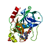
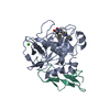
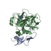
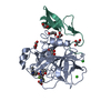
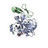
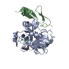
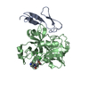
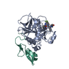
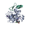
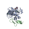
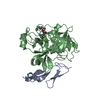
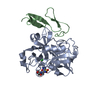
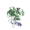
| Title | Crystal Structure of Human Coagulation Factor XA Complexed with RPR208707 | ||||||
 Components Components | (COAGULATION FACTOR XA) x 2 | ||||||
 Keywords Keywords | HYDROLASE / Protein-inhibitor complex | ||||||
| Function / homology |  Function and homology information Function and homology informationcoagulation factor Xa / Defective factor IX causes thrombophilia / Defective cofactor function of FVIIIa variant / Defective F9 variant does not activate FX / : / positive regulation of TOR signaling / Transport of gamma-carboxylated protein precursors from the endoplasmic reticulum to the Golgi apparatus / : / Gamma-carboxylation of protein precursors / Removal of aminoterminal propeptides from gamma-carboxylated proteins ...coagulation factor Xa / Defective factor IX causes thrombophilia / Defective cofactor function of FVIIIa variant / Defective F9 variant does not activate FX / : / positive regulation of TOR signaling / Transport of gamma-carboxylated protein precursors from the endoplasmic reticulum to the Golgi apparatus / : / Gamma-carboxylation of protein precursors / Removal of aminoterminal propeptides from gamma-carboxylated proteins / : / phospholipid binding / Golgi lumen / blood coagulation / positive regulation of cell migration / endoplasmic reticulum lumen / serine-type endopeptidase activity / external side of plasma membrane / calcium ion binding / proteolysis / : / extracellular region / plasma membrane Similarity search - Function | ||||||
| Biological species |  Homo sapiens (human) Homo sapiens (human) | ||||||
| Method |  X-RAY DIFFRACTION / Resolution: 2.1 Å X-RAY DIFFRACTION / Resolution: 2.1 Å | ||||||
 Authors Authors | Maignan, S. / Guilloteau, J.P. / Pouzieux, S. / Choi-Sledeski, Y.M. / Becker, M.R. / Klein, S.I. / Ewing, W.R. / Pauls, H.W. / Spada, A.P. / Mikol, V. | ||||||
 Citation Citation | Journal: J.Med.Chem. / Year: 2000 Title: Crystal structures of human factor Xa complexed with potent inhibitors. Authors: Maignan, S. / Guilloteau, J.P. / Pouzieux, S. / Choi-Sledeski, Y.M. / Becker, M.R. / Klein, S.I. / Ewing, W.R. / Pauls, H.W. / Spada, A.P. / Mikol, V. | ||||||
| History |
|
- Structure visualization
| Structure viewer | Molecule: MolmilJmol/JSmol |
|---|
- Downloads & links
-Download
| PDBx/mmCIF format | 1f0s.cif.gz | 76.9 KB | Display | PDBx/mmCIF format |
|---|---|---|---|---|
| PDB format | pdb1f0s.ent.gz | 54.5 KB | Display | PDB format |
| PDBx/mmJSON format | 1f0s.json.gz | Tree view | PDBx/mmJSON format | |
| Others |  Other downloads Other downloads |
-Validation report
| Arichive directory | https://data.pdbj.org/pub/pdb/validation_reports/f0/1f0sftp://data.pdbj.org/pub/pdb/validation_reports/f0/1f0s | HTTPS FTP |
|---|
-Related structure data


-Links
 PDBj
PDBj


- Assembly
| Deposited unit | 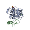
| ||||||||
|---|---|---|---|---|---|---|---|---|---|
| 1 |
| ||||||||
| Unit cell |
| ||||||||
| Details | The biological assembly is constructed from chain A and B linked through a disulfide bridge |
-Components
| #1: Protein | Mass: 28550.596 Da / Num. of mol.: 1 / Fragment: ACTIVATED FACTOR XA, HEAVY CHAIN / Source method: isolated from a natural source / Source: (natural) Homo sapiens (human) / References: UniProt: P00742, coagulation factor Xa |
|---|---|
| #2: Protein | Mass: 15210.793 Da / Num. of mol.: 1 / Fragment: FACTOR X LIGHT CHAIN / Source method: isolated from a natural source / Source: (natural) Homo sapiens (human) / References: UniProt: P00742, coagulation factor Xa |
| #3: Chemical | ChemComp-CA /   Mass: 40.078 Da / Num. of mol.: 1 / Source method: obtained synthetically / Formula: Ca / Details: chemical synthesis Mass: 40.078 Da / Num. of mol.: 1 / Source method: obtained synthetically / Formula: Ca / Details: chemical synthesis |
| #4: Chemical | ChemComp-PR2 /   Mass: 427.500 Da / Num. of mol.: 1 / Source method: obtained synthetically / Formula: C19H17N5O3S2 Mass: 427.500 Da / Num. of mol.: 1 / Source method: obtained synthetically / Formula: C19H17N5O3S2 |
| #5: Water | ChemComp-HOH /  Mass: 18.015 Da / Num. of mol.: 147 / Source method: isolated from a natural source / Formula: H2O Mass: 18.015 Da / Num. of mol.: 147 / Source method: isolated from a natural source / Formula: H2O |
| Has protein modification | Y |
| Sequence details | Some residues in chains A and B are not seen in the electron density. Sequence database Residues 1- ...Some residues in chains A and B are not seen in the electron density. Sequence database Residues 1-45 (the Gla domain) were biochemically removed in chain B. |
-Experimental details
-Experiment
| Experiment | Method: X-RAY DIFFRACTION / Number of used crystals: 1 |
|---|
- Sample preparation
| Crystal grow | Temperature: 292 K / Method: vapor diffusion, hanging drop / pH: 5.7 Details: 18-22%PEG 600, 50mM Mes-NaOH, pH 5.7, VAPOR DIFFUSION, HANGING DROP, temperature 292K | ||||||||||||||||||||||||||||||||||||||||||
|---|---|---|---|---|---|---|---|---|---|---|---|---|---|---|---|---|---|---|---|---|---|---|---|---|---|---|---|---|---|---|---|---|---|---|---|---|---|---|---|---|---|---|---|
| Crystal grow | *PLUS Temperature: 19 ℃ / pH: 6 | ||||||||||||||||||||||||||||||||||||||||||
| Components of the solutions | *PLUS
|
-Data collection
| Diffraction | Mean temperature: 95 K |
|---|---|
| Diffraction source | Source: ROTATING ANODE / Type: ENRAF-NONIUS FR591 / Wavelength: 1.5418 / Wavelength: 1.5418 Å |
| Detector | Type: MAC Science DIP-2000 / Detector: IMAGE PLATE / Date: Mar 7, 1998 |
| Radiation | Protocol: SINGLE WAVELENGTH / Monochromatic (M) / Laue (L): M / Scattering type: x-ray |
| Radiation wavelength | Wavelength: 1.5418 Å / Relative weight: 1 |
| Reflection | Resolution: 2.1→20 Å / Num. all: 17166 / Num. obs: 17166 / % possible obs: 90.5 % / Observed criterion σ(F): 1 / Observed criterion σ(I): 1 / Rmerge(I) obs: 0.084 |
| Reflection shell | Resolution: 2.1→2.17 Å / Rmerge(I) obs: 0.192 / % possible all: 68.8 |
| Reflection | *PLUS |
| Reflection shell | *PLUS % possible obs: 68.6 % / Redundancy: 3.1 % / Rmerge(I) obs: 0.217 |
- Processing
| Software |
| ||||||||||||||||||||
|---|---|---|---|---|---|---|---|---|---|---|---|---|---|---|---|---|---|---|---|---|---|
| Refinement | Resolution: 2.1→20 Å / σ(F): 0 / σ(I): 0 / Stereochemistry target values: Engh & Huber
| ||||||||||||||||||||
| Refinement step | Cycle: LAST / Resolution: 2.1→20 Å
| ||||||||||||||||||||
| Refine LS restraints |
| ||||||||||||||||||||
| Software | *PLUS Name: X-PLOR / Version: 98 / Classification: refinement | ||||||||||||||||||||
| Refinement | *PLUS | ||||||||||||||||||||
| Solvent computation | *PLUS | ||||||||||||||||||||
| Displacement parameters | *PLUS | ||||||||||||||||||||
| Refine LS restraints | *PLUS
|
