 Movie
Movie Controller

[English] 日本語
 Yorodumi
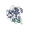
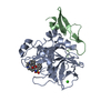
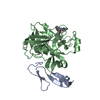
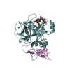
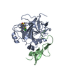
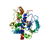
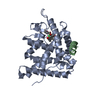
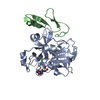
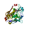
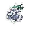
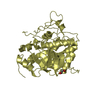
- PDB-1nfu: CRYSTAL STRUCTURE OF HUMAN COAGULATION FACTOR XA COMPLEXED WITH R... -
+ Open data

- Basic information
| Entry | Database: PDB / ID: 1nfu | ||||||
|---|---|---|---|---|---|---|---|
| Title | CRYSTAL STRUCTURE OF HUMAN COAGULATION FACTOR XA COMPLEXED WITH RPR132747 | ||||||
 Components Components |
| ||||||
 Keywords Keywords | HYDROLASE | ||||||
| Function / homology |  Function and homology information Function and homology informationcoagulation factor Xa / Defective factor IX causes thrombophilia / Defective cofactor function of FVIIIa variant / Defective F9 variant does not activate FX / : / positive regulation of TOR signaling / Transport of gamma-carboxylated protein precursors from the endoplasmic reticulum to the Golgi apparatus / : / Gamma-carboxylation of protein precursors / Removal of aminoterminal propeptides from gamma-carboxylated proteins ...coagulation factor Xa / Defective factor IX causes thrombophilia / Defective cofactor function of FVIIIa variant / Defective F9 variant does not activate FX / : / positive regulation of TOR signaling / Transport of gamma-carboxylated protein precursors from the endoplasmic reticulum to the Golgi apparatus / : / Gamma-carboxylation of protein precursors / Removal of aminoterminal propeptides from gamma-carboxylated proteins / : / phospholipid binding / Golgi lumen / blood coagulation / positive regulation of cell migration / endoplasmic reticulum lumen / serine-type endopeptidase activity / external side of plasma membrane / calcium ion binding / proteolysis / : / extracellular region / plasma membrane Similarity search - Function | ||||||
| Biological species |  Homo sapiens (human) Homo sapiens (human) | ||||||
| Method |  X-RAY DIFFRACTION / SYNCHROTRON / MOLECULAR REPLACEMENT / Resolution: 2.05 Å X-RAY DIFFRACTION / SYNCHROTRON / MOLECULAR REPLACEMENT / Resolution: 2.05 Å | ||||||
 Authors Authors | Maignan, S. / Guilloteau, J.P. | ||||||
 Citation Citation | Journal: J.Med.Chem. / Year: 2003 Title: Molecular structures of human Factor Xa complexed with ketopiperazine inhibitors: preference for a neutral group in the S1 pocket. Authors: Maignan, S. / Guilloteau, J.P. / Choi-Sledeski, Y.M. / Becker, M.R. / Ewing, W.R. / Pauls, H.W. / Spada, A.P. / Mikol, V. | ||||||
| History |
|
- Structure visualization
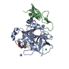
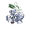
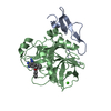
| Structure viewer | Molecule: MolmilJmol/JSmol |
|---|
- Downloads & links
-Download
| PDBx/mmCIF format | 1nfu.cif.gz | 79.3 KB | Display | PDBx/mmCIF format |
|---|---|---|---|---|
| PDB format | pdb1nfu.ent.gz | 55.6 KB | Display | PDB format |
| PDBx/mmJSON format | 1nfu.json.gz | Tree view | PDBx/mmJSON format | |
| Others |  Other downloads Other downloads |
-Validation report
| Arichive directory | https://data.pdbj.org/pub/pdb/validation_reports/nf/1nfuftp://data.pdbj.org/pub/pdb/validation_reports/nf/1nfu | HTTPS FTP |
|---|
-Related structure data
| Related structure data |  1nfwC  1nfxC  1nfyC  1fosS C: citing same article ( S: Starting model for refinement |
|---|---|
| Similar structure data |


-Links
 PDBj
PDBj


- Assembly
| Deposited unit | 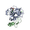
| ||||||||
|---|---|---|---|---|---|---|---|---|---|
| 1 |
| ||||||||
| Unit cell |
|
-Components
| #1: Protein | Mass: 28550.596 Da / Num. of mol.: 1 / Source method: isolated from a natural source / Details: Blood / Source: (natural) Homo sapiens (human) / References: UniProt: P00742, coagulation factor Xa |
|---|---|
| #2: Protein | Mass: 21919.961 Da / Num. of mol.: 1 / Source method: isolated from a natural source / Details: Blood / Source: (natural) Homo sapiens (human) / References: UniProt: P00742, coagulation factor Xa |
| #3: Chemical | ChemComp-CA /   Mass: 40.078 Da / Num. of mol.: 1 / Source method: obtained synthetically / Formula: Ca Mass: 40.078 Da / Num. of mol.: 1 / Source method: obtained synthetically / Formula: Ca |
| #4: Chemical | ChemComp-RRP / 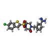  Mass: 462.973 Da / Num. of mol.: 1 / Source method: obtained synthetically / Formula: C20H19ClN4O3S2 Mass: 462.973 Da / Num. of mol.: 1 / Source method: obtained synthetically / Formula: C20H19ClN4O3S2 |
| #5: Water | ChemComp-HOH /  Mass: 18.015 Da / Num. of mol.: 163 / Source method: isolated from a natural source / Formula: H2O Mass: 18.015 Da / Num. of mol.: 163 / Source method: isolated from a natural source / Formula: H2O |
| Has protein modification | Y |
-Experimental details
-Experiment
| Experiment | Method: X-RAY DIFFRACTION / Number of used crystals: 1 |
|---|
- Sample preparation
| Crystal grow | Temperature: 292 K / Method: vapor diffusion, hanging drop / pH: 5.7 Details: 18-20% PEG 600, 50MM MES-NAOH,1MM RPR132747, pH 5.7, VAPOR DIFFUSION, HANGING DROP, temperature 292K | |||||||||||||||||||||||||||||||||||||||||||||||||
|---|---|---|---|---|---|---|---|---|---|---|---|---|---|---|---|---|---|---|---|---|---|---|---|---|---|---|---|---|---|---|---|---|---|---|---|---|---|---|---|---|---|---|---|---|---|---|---|---|---|---|
| Crystal grow | *PLUS Temperature: 19 ℃ / pH: 6 | |||||||||||||||||||||||||||||||||||||||||||||||||
| Components of the solutions | *PLUS
|
-Data collection
| Diffraction | Mean temperature: 100 K |
|---|---|
| Diffraction source | Source: SYNCHROTRON / Site: ESRF  / Beamline: ID14-3 / Wavelength: 0.933 Å / Beamline: ID14-3 / Wavelength: 0.933 Å |
| Detector | Type: MARRESEARCH / Detector: CCD / Date: Aug 1, 1999 |
| Radiation | Protocol: SINGLE WAVELENGTH / Monochromatic (M) / Laue (L): M / Scattering type: x-ray |
| Radiation wavelength | Wavelength: 0.933 Å / Relative weight: 1 |
| Reflection | Resolution: 2.05→26 Å / Num. all: 18808 / Num. obs: 18808 / % possible obs: 92 % / Observed criterion σ(F): 0 / Observed criterion σ(I): 1 / Rmerge(I) obs: 0.032 |
| Reflection | *PLUS Redundancy: 2.4 % |
| Reflection shell | *PLUS % possible obs: 89.4 % / Rmerge(I) obs: 0.118 |
- Processing
| Software |
| |||||||||||||||
|---|---|---|---|---|---|---|---|---|---|---|---|---|---|---|---|---|
| Refinement | Method to determine structure: MOLECULAR REPLACEMENT Starting model: PDB ENTRY 1FOS Resolution: 2.05→20 Å / σ(F): 0 / Stereochemistry target values: Engh & Huber Details: refinement of the structure was performed without r-free calculation
| |||||||||||||||
| Displacement parameters | Biso mean: 29.7 Å2 | |||||||||||||||
| Refinement step | Cycle: LAST / Resolution: 2.05→20 Å
| |||||||||||||||
| Refinement | *PLUS | |||||||||||||||
| Solvent computation | *PLUS | |||||||||||||||
| Displacement parameters | *PLUS | |||||||||||||||
| Refine LS restraints | *PLUS
|
