 Movie
Movie Controller

[English] 日本語
 Yorodumi
- PDB-2xxm: Crystal structure of the HIV-1 capsid protein C-terminal domain i... -
+ Open data

- Basic information
| Entry | Database: PDB / ID: 2xxm | ||||||
|---|---|---|---|---|---|---|---|
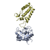
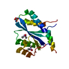
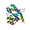
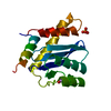
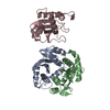
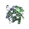
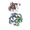
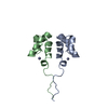
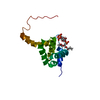
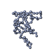
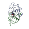
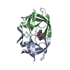
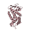
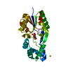
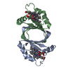
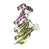
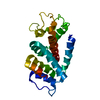
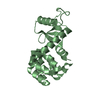
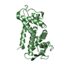
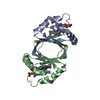
| Title | Crystal structure of the HIV-1 capsid protein C-terminal domain in complex with a camelid VHH and the CAI peptide. | ||||||
 Components Components |
| ||||||
 Keywords Keywords | IMMUNE SYSTEM/PEPTIDE / IMMUNE SYSTEM-PEPTIDE COMPLEX / CAPSID INHIBITOR / PROTEIN BINDING / PROTEIN INTERFACE / VIRUS ASSEMBLY | ||||||
| Function / homology |  Function and homology information Function and homology informationHIV-1 retropepsin / symbiont-mediated activation of host apoptosis / retroviral ribonuclease H / exoribonuclease H / exoribonuclease H activity / DNA integration / viral genome integration into host DNA / establishment of integrated proviral latency / RNA-directed DNA polymerase / RNA stem-loop binding ...HIV-1 retropepsin / symbiont-mediated activation of host apoptosis / retroviral ribonuclease H / exoribonuclease H / exoribonuclease H activity / DNA integration / viral genome integration into host DNA / establishment of integrated proviral latency / RNA-directed DNA polymerase / RNA stem-loop binding / viral penetration into host nucleus / host multivesicular body / RNA-directed DNA polymerase activity / RNA-DNA hybrid ribonuclease activity / Transferases; Transferring phosphorus-containing groups; Nucleotidyltransferases / host cell / viral nucleocapsid / DNA recombination / DNA-directed DNA polymerase / aspartic-type endopeptidase activity / Hydrolases; Acting on ester bonds / DNA-directed DNA polymerase activity / symbiont-mediated suppression of host gene expression / viral translational frameshifting / symbiont entry into host cell / lipid binding / host cell nucleus / host cell plasma membrane / virion membrane / structural molecule activity / proteolysis / DNA binding / zinc ion binding Similarity search - Function | ||||||
| Biological species |   HUMAN IMMUNODEFICIENCY VIRUS HUMAN IMMUNODEFICIENCY VIRUS SYNTHETIC CONSTRUCT (others) | ||||||
| Method |  X-RAY DIFFRACTION / SYNCHROTRON / MOLECULAR REPLACEMENT / Resolution: 1.65 Å X-RAY DIFFRACTION / SYNCHROTRON / MOLECULAR REPLACEMENT / Resolution: 1.65 Å | ||||||
 Authors Authors | Igonet, S. / Vaney, M.C. / Bartonova, V. / Helma, J. / Rothbauer, U. / Leonhardt, H. / Stura, E. / Krausslich, H.-G. / Rey, F.A. | ||||||
 Citation Citation | Journal: To be Published Title: Targeting HIV-1 Virion Formation with Nanobodies -Implications for the Design of Assembly Inhibitors Authors: Igonet, S. / Vaney, M.C. / Bartonova, V. / Helma, J. / Rothbauer, U. / Leonhardt, H. / Stura, E. / Krausslich, H.-G. / Rey, F.A. | ||||||
| History |
|
- Structure visualization
| Structure viewer | Molecule: MolmilJmol/JSmol |
|---|
- Downloads & links
-Download
| PDBx/mmCIF format | 2xxm.cif.gz | 56.6 KB | Display | PDBx/mmCIF format |
|---|---|---|---|---|
| PDB format | pdb2xxm.ent.gz | 40.3 KB | Display | PDB format |
| PDBx/mmJSON format | 2xxm.json.gz | Tree view | PDBx/mmJSON format | |
| Others |  Other downloads Other downloads |
-Validation report
| Arichive directory | https://data.pdbj.org/pub/pdb/validation_reports/xx/2xxmftp://data.pdbj.org/pub/pdb/validation_reports/xx/2xxm | HTTPS FTP |
|---|
-Related structure data
| Related structure data |  2xt1C  2xv6SC 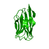 2xxcC C: citing same article ( S: Starting model for refinement |
|---|---|
| Similar structure data |


-Links
 PDBj
PDBj


- Assembly
| Deposited unit | 
| |||||||||
|---|---|---|---|---|---|---|---|---|---|---|
| 1 |
| |||||||||
| Unit cell |
| |||||||||
| Components on special symmetry positions |
|
-Components
| #1: Protein | Mass: 8455.631 Da / Num. of mol.: 1 / Fragment: C-TERMINAL DOMAIN, RESIDUES 278-352 Source method: isolated from a genetically manipulated source Source: (gene. exp.) HUMAN IMMUNODEFICIENCY VIRUS / Strain: NL4-3 / Plasmid: PET11C / Production host:  | ||||
|---|---|---|---|---|---|
| #2: Antibody | Mass: 13213.812 Da / Num. of mol.: 1 Source method: isolated from a genetically manipulated source Source: (gene. exp.) | ||||
| #3: Protein/peptide | Mass: 1348.455 Da / Num. of mol.: 1 / Source method: obtained synthetically / Source: (synth.) SYNTHETIC CONSTRUCT (others) | ||||
| #4: Chemical |   Mass: 59.044 Da / Num. of mol.: 3 / Source method: obtained synthetically / Formula: C2H3O2 Mass: 59.044 Da / Num. of mol.: 3 / Source method: obtained synthetically / Formula: C2H3O2#5: Water | ChemComp-HOH / |  Mass: 18.015 Da / Num. of mol.: 180 / Source method: isolated from a natural source / Formula: H2O Mass: 18.015 Da / Num. of mol.: 180 / Source method: isolated from a natural source / Formula: H2OHas protein modification | Y | |
-Experimental details
-Experiment
| Experiment | Method: X-RAY DIFFRACTION / Number of used crystals: 5 |
|---|
- Sample preparation
| Crystal | Density Matthews: 2.03 Å3/Da / Density % sol: 39.4 % / Description: NONE |
|---|---|
| Crystal grow | Details: 30% PEG4000, 200MM AMMONIUM ACETATE, 100MM SODIUM ACETATE PH 4.6 |
-Data collection
| Diffraction | Mean temperature: 100 K |
|---|---|
| Diffraction source | Source: SYNCHROTRON / Site: SLS  / Beamline: X06SA / Wavelength: 0.98 / Beamline: X06SA / Wavelength: 0.98 |
| Detector | Type: DECTRIS PILATUS 6M / Detector: PIXEL / Date: Feb 21, 2009 / Details: DYNAMICALLY BENDABLE MIRROR |
| Radiation | Monochromator: SI(111) MONOCHROMATOR / Protocol: SINGLE WAVELENGTH / Monochromatic (M) / Laue (L): M / Scattering type: x-ray |
| Radiation wavelength | Wavelength: 0.98 Å / Relative weight: 1 |
| Reflection | Resolution: 1.65→40 Å / Num. obs: 22400 / % possible obs: 95.4 % / Observed criterion σ(I): 0 / Redundancy: 5.5 % / Biso Wilson estimate: 19.96 Å2 / Rmerge(I) obs: 0.09 / Net I/σ(I): 13.1 |
| Reflection shell | Resolution: 1.65→1.74 Å / Redundancy: 1.9 % / Rmerge(I) obs: 0.36 / Mean I/σ(I) obs: 2 / % possible all: 77.8 |
- Processing
| Software |
| ||||||||||||||||||||||||||||||||||||||||||||||||||||||||||||||||||||||||||||||||||||||||||||||||||||||||||||||||||
|---|---|---|---|---|---|---|---|---|---|---|---|---|---|---|---|---|---|---|---|---|---|---|---|---|---|---|---|---|---|---|---|---|---|---|---|---|---|---|---|---|---|---|---|---|---|---|---|---|---|---|---|---|---|---|---|---|---|---|---|---|---|---|---|---|---|---|---|---|---|---|---|---|---|---|---|---|---|---|---|---|---|---|---|---|---|---|---|---|---|---|---|---|---|---|---|---|---|---|---|---|---|---|---|---|---|---|---|---|---|---|---|---|---|---|---|
| Refinement | Method to determine structure: MOLECULAR REPLACEMENT Starting model: PDB ENTRY 2XV6 Resolution: 1.65→37.89 Å / Cor.coef. Fo:Fc: 0.9453 / Cor.coef. Fo:Fc free: 0.9427 / SU R Cruickshank DPI: 0.106 / Cross valid method: THROUGHOUT / σ(F): 0 / SU R Blow DPI: 0.116 / SU Rfree Blow DPI: 0.105 / SU Rfree Cruickshank DPI: 0.1
| ||||||||||||||||||||||||||||||||||||||||||||||||||||||||||||||||||||||||||||||||||||||||||||||||||||||||||||||||||
| Displacement parameters | Biso mean: 22.42 Å2
| ||||||||||||||||||||||||||||||||||||||||||||||||||||||||||||||||||||||||||||||||||||||||||||||||||||||||||||||||||
| Refine analyze | Luzzati coordinate error obs: 0.193 Å | ||||||||||||||||||||||||||||||||||||||||||||||||||||||||||||||||||||||||||||||||||||||||||||||||||||||||||||||||||
| Refinement step | Cycle: LAST / Resolution: 1.65→37.89 Å
| ||||||||||||||||||||||||||||||||||||||||||||||||||||||||||||||||||||||||||||||||||||||||||||||||||||||||||||||||||
| Refine LS restraints |
| ||||||||||||||||||||||||||||||||||||||||||||||||||||||||||||||||||||||||||||||||||||||||||||||||||||||||||||||||||
| LS refinement shell | Resolution: 1.65→1.73 Å / Total num. of bins used: 11
|
